. 2021 Sep;42(13):4081-4091.
doi: 10.1002/hbm.24497. Epub 2019 Jan 3.

MR-PET head motion correction based on co-registration of multicontrast MR images

Affiliations

MR-PET head motion correction based on co-registration of multicontrast MR images

Zhaolin Chen et al. Hum Brain Mapp. 2021 Sep.

Abstract

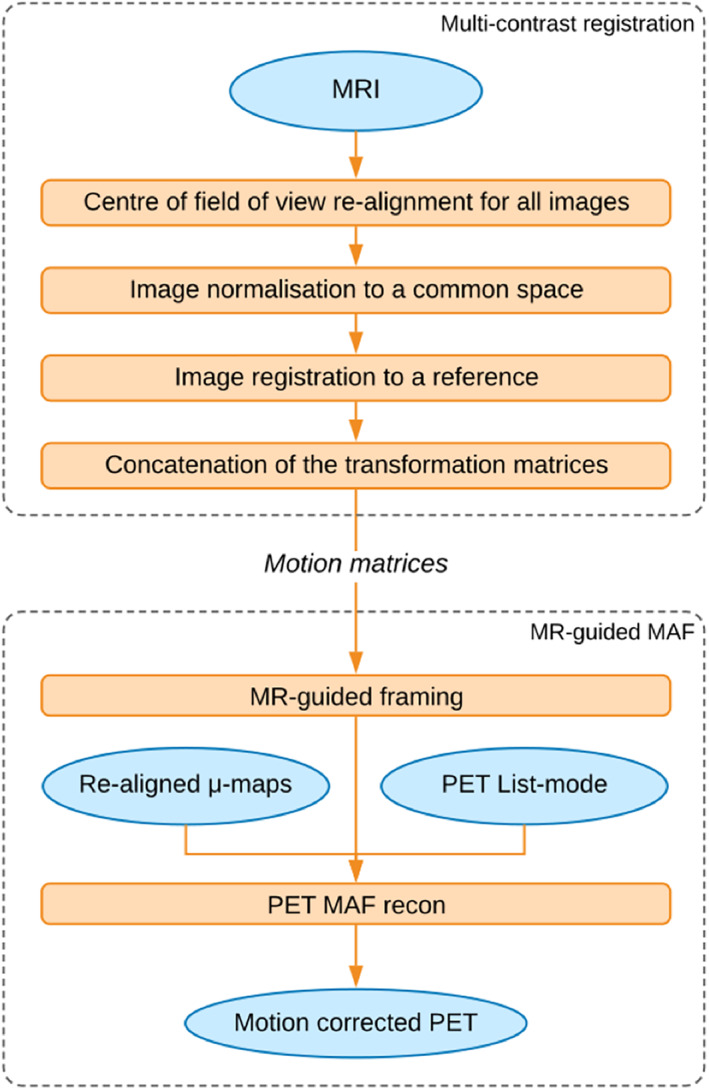
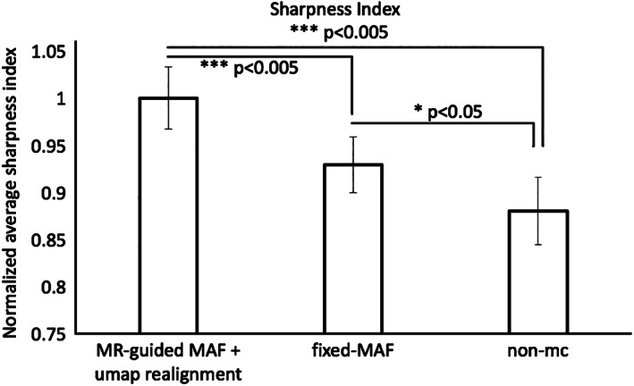
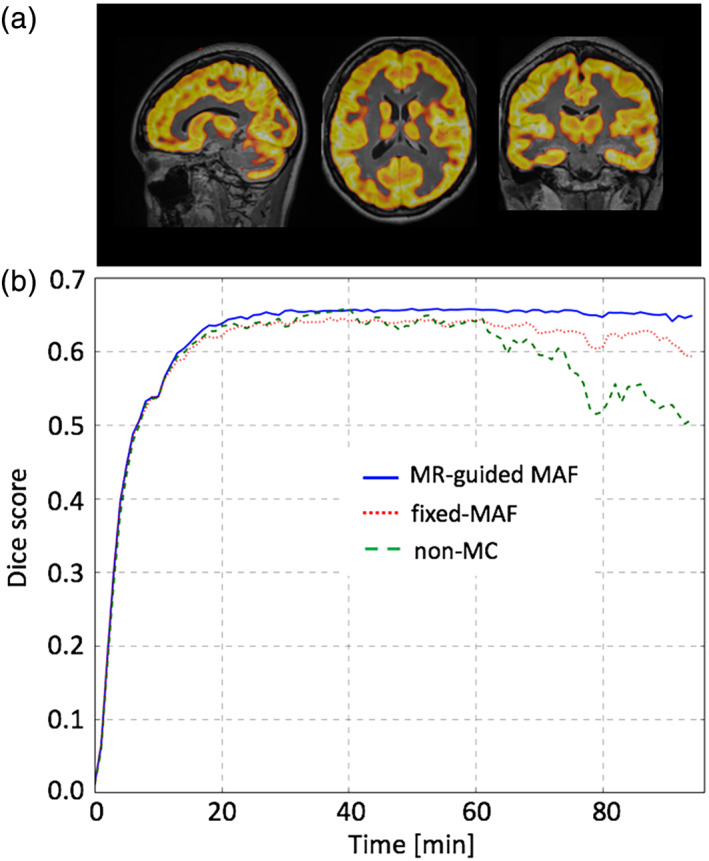
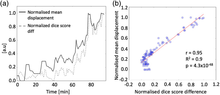
Head motion is a major source of image artefacts in neuroimaging studies and can lead to degradation of the quantitative accuracy of reconstructed PET images. Simultaneous magnetic resonance-positron emission tomography (MR-PET) makes it possible to estimate head motion information from high-resolution MR images and then correct motion artefacts in PET images. In this article, we introduce a fully automated PET motion correction method, MR-guided MAF, based on the co-registration of multicontrast MR images. The performance of the MR-guided MAF method was evaluated using MR-PET data acquired from a cohort of ten healthy participants who received a slow infusion of fluorodeoxyglucose ([18-F]FDG). Compared with conventional methods, MR-guided PET image reconstruction can reduce head motion introduced artefacts and improve the image sharpness and quantitative accuracy of PET images acquired using simultaneous MR-PET scanners. The fully automated motion estimation method has been implemented as a publicly available web-service.

Keywords: MR image registration; MR-guided MAF; MR-guided motion correction; PET motion artefacts; PET motion correction; PET/MR; multiple acquisition frame (MAF); simultaneous MR-PET.


Conflict of interest statement

None.

Figures

FIGURE 1
FIGURE 1
Overview of the MR‐guided MAF method
FIGURE 2
FIGURE 2
Mean displacement plot for the motion instructed volunteer demonstrating the head movement with respect to the T2 weighted reference image, as detected by the multicontrast registration method. The yellow/white alternation bands indicate the durations of successive motion correction frames
FIGURE 3
FIGURE 3
Motion correction results for the controlled motion experiment. Images in panels (a)–(d) show the reconstructed PET images using different reconstruction methods. The plots in panel (e) show the signal intensity variation along the line profiles in panel (a)–(d)
FIGURE 4
FIGURE 4
Comparison of the motion correction results for the group averaged image. The images in panels (a)–(c) show the reconstructed PET images using the three different reconstruction methods. The plots in panel (d) show the signal intensity variation along the line profiles in panels (a)–(c)
FIGURE 5
FIGURE 5
Comparison of the averaged (mean and standard errors) sharpness indices for the experimental group of 10 participants
FIGURE 6
FIGURE 6
Comparison of the motion correction results between the MR‐guided MAF, fixed‐MAF and for the images without motion correction, for a dynamic PET reconstruction for one test subject. The Dice scores are shown in (b) using grey matter masks shown in (a)
FIGURE 7
FIGURE 7
Plots of the Dice score differences and the mean displacement between the MR‐based motion corrected and the nonmotion corrected PET images in panel (a). Panel (b) shows the correlation scatter plot between the Dice score differences and the mean displacement

References

    1. Alexander‐Bloch, A., Clasen, L., Stockman, M., Ronan, L., Lalonde, F., Giedd, J., & Raznahan, A. (2016). Subtle in‐scanner motion biases automated measurement of brain anatomy from in vivo MRI. Human Brain Mapping, 37, 2385–2397. 10.1002/hbm.23180 - DOI - PMC - PubMed
    1. Andersson, J. L. R., Skare, S., & Ashburner, J. (2003). How to correct susceptibility distortions in spin‐echo echo‐planar images: Application to diffusion tensor imaging. NeuroImage, 20, 870–888. 10.1016/S1053-8119(03)00336-7 - DOI - PubMed
    1. Andersson, J. L. R., & Sotiropoulos, S. N. (2016). An integrated approach to correction for off‐resonance effects and subject movement in diffusion MR imaging. NeuroImage, 125, 1063–1078. 10.1016/j.neuroimage.2015.10.019 - DOI - PMC - PubMed
    1. Ardekani, B. A., Bachman, A. H., & Helpern, J. A. (2001). A quantitative comparison of motion detection algorithms in fMRI. Magnetic Resonance Imaging, 19, 959–963. - PubMed
    1. Baran, J., Chen, Z., Sforazzini, F., Ferris, N., Jamadar, S., Schmitt, B., … Egan, G. F. (2018). Accurate hybrid template‐based and MR‐based attenuation correction using UTE images for simultaneous PET/MR brain imaging applications. BMC Medical Imaging, 18, 41. - PMC - PubMed

Publication types