

Amyloid burden accelerates white matter degradation in cognitively normal elderly individuals
- PMID: 30604903
- PMCID: PMC6501192
- DOI: 10.1002/hbm.24507
Amyloid burden accelerates white matter degradation in cognitively normal elderly individuals
Abstract
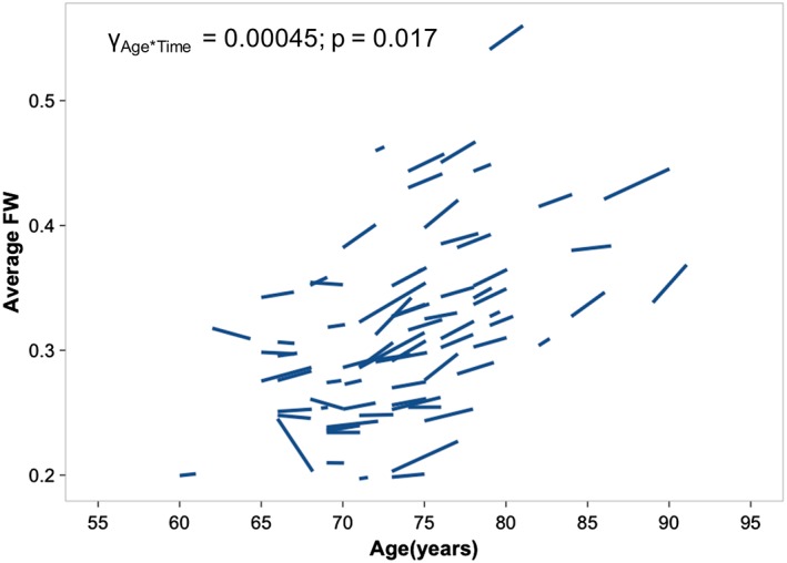
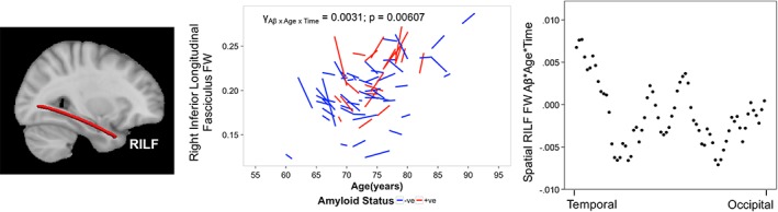
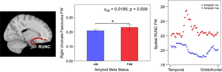
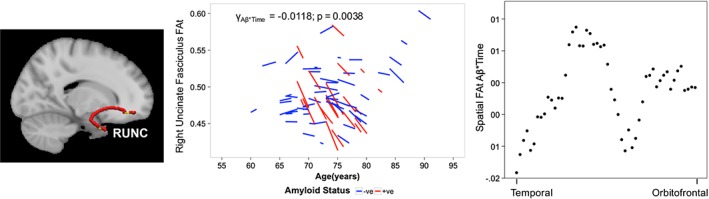
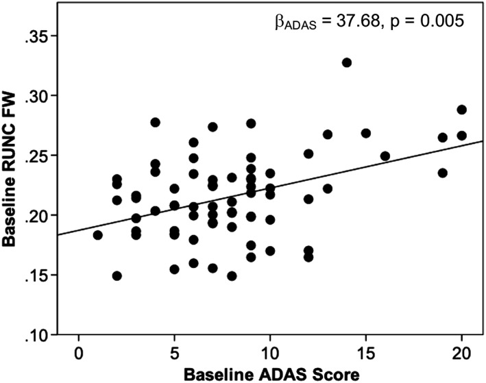
Alterations in parietal and temporal white matter microstructure derived from diffusion tensor imaging occur in preclinical and clinical Alzheimer's disease. Amyloid beta (Aβ) deposition and such white matter alterations are two pathological hallmarks of Alzheimer's disease. However, the relationship between these pathologies is not yet understood, partly since conventional diffusion MRI methods cannot distinguish between cellular and extracellular processes. Thus, we studied Aβ-associated longitudinal diffusion MRI changes in Aβ-positive (N = 21) and Aβ-negative (N = 51) cognitively normal elderly obtained from the Alzheimer's Disease Neuroimaging Initiative dataset using linear mixed models. Aβ-positivity was based on Alzheimer's Disease Neuroimaging Initiative amyloid-PET recommendations using a standardized uptake value ratio cut-off of 1.11. We used free-water imaging to distinguish cellular and extracellular changes. We found that Aβ-positive subjects had increased baseline right uncinate fasciculus free-water fraction (FW), associated with worse baseline Alzheimer's disease assessment scale scores. Furthermore, Aβ-positive subjects showed faster decrease in fractional anisotropy (FW-corrected) in the right uncinate fasciculus and faster age-dependent right inferior longitudinal fasciculus FW increases over time. Right inferior longitudinal fasciculus FW increases were associated with greater memory decline. Importantly, these results remained significant after controlling for gray and white matter volume and hippocampal volume. This is the first study to illustrate the influence of Aβ burden on early longitudinal (in addition to baseline) white matter changes in cognitively normal elderly individuals at-risk of Alzheimer's disease, thus underscoring the importance of longitudinal studies in assessing microstructural alterations in individuals at risk of Alzheimer's disease prior to symptoms onset.
Keywords: Alzheimer's disease; amyloid beta; at-risk cognitively normal elderly; diffusion tensor imaging; free-water correction; longitudinal study.
© 2019 Wiley Periodicals, Inc.
Conflict of interest statement
The authors have no potential conflicts of interest.
Figures
Similar articles
-
Associations between white matter microstructure and amyloid burden in preclinical Alzheimer's disease: A multimodal imaging investigation.Neuroimage Clin. 2014 Feb 19;4:604-14. doi: 10.1016/j.nicl.2014.02.001. eCollection 2014. Neuroimage Clin. 2014. PMID: 24936411 Free PMC article.
-
Free Water MR Imaging of White Matter Microstructural Changes is a Sensitive Marker of Amyloid Positivity in Alzheimer's Disease.J Magn Reson Imaging. 2024 Oct;60(4):1458-1469. doi: 10.1002/jmri.29189. Epub 2023 Dec 15. J Magn Reson Imaging. 2024. PMID: 38100518
-
White matter changes in preclinical Alzheimer's disease: a magnetic resonance imaging-diffusion tensor imaging study on cognitively normal older people with positive amyloid β protein 42 levels.Neurobiol Aging. 2014 Dec;35(12):2671-2680. doi: 10.1016/j.neurobiolaging.2014.05.027. Epub 2014 Jun 6. Neurobiol Aging. 2014. PMID: 25002037
-
White matter hyperintensities are higher among early-onset Alzheimer's disease participants than their cognitively normal and early-onset nonAD peers: Longitudinal Early-onset Alzheimer's Disease Study (LEADS).Alzheimers Dement. 2023 Nov;19 Suppl 9(Suppl 9):S89-S97. doi: 10.1002/alz.13402. Epub 2023 Jul 25. Alzheimers Dement. 2023. PMID: 37491599 Free PMC article. Review.
-
Diffusion tensor imaging of white matter degeneration in early stage of Alzheimer's disease: a review.Int J Neurosci. 2020 Mar;130(3):243-250. doi: 10.1080/00207454.2019.1667798. Epub 2019 Sep 24. Int J Neurosci. 2020. PMID: 31549530
Cited by
-
Tract-specific differences in white matter microstructure between young adult APOE ε4 carriers and non-carriers: A replication and extension study.Neuroimage Rep. 2022 Dec;2(4):None. doi: 10.1016/j.ynirp.2022.100126. Neuroimage Rep. 2022. PMID: 36507069 Free PMC article.
-
Improving brain age prediction models: incorporation of amyloid status in Alzheimer's disease.Neurobiol Aging. 2020 Mar;87:44-48. doi: 10.1016/j.neurobiolaging.2019.11.005. Epub 2019 Nov 14. Neurobiol Aging. 2020. PMID: 31843257 Free PMC article.
-
Searching for optimal machine learning model to classify mild cognitive impairment (MCI) subtypes using multimodal MRI data.Sci Rep. 2022 Mar 11;12(1):4284. doi: 10.1038/s41598-022-08231-y. Sci Rep. 2022. PMID: 35277565 Free PMC article.
-
Heart-brain mapping: Cardiac atrial function is associated with distinct cerebral regions with high free water in older adults.J Cereb Blood Flow Metab. 2024 Jul;44(7):1218-1230. doi: 10.1177/0271678X241229581. Epub 2024 Jan 31. J Cereb Blood Flow Metab. 2024. PMID: 38295860 Free PMC article.
-
Small vessel disease more than Alzheimer's disease determines diffusion MRI alterations in memory clinic patients.Alzheimers Dement. 2020 Nov;16(11):1504-1514. doi: 10.1002/alz.12150. Epub 2020 Aug 18. Alzheimers Dement. 2020. PMID: 32808747 Free PMC article.
References
-
- ADNI . (2011). ADNI 2 PET Technical Procedures Manual AV‐45 (Florbetapir F 18) & FDG. Boston, MA: ADNI.
-
- Alzheimer Disease Neuroimaging Initiative . (2008). ADNI 2 Procedures Manual. Boston, MA: ADNI.
-
- Ashtari, M. (2012). Anatomy and functional role of the inferior longitudinal fasciculus: A search that has just begun. Developmental Medicine and Child Neurology, 54, 6–7. - PubMed
Publication types
MeSH terms
Substances
Grants and funding
LinkOut - more resources
Full Text Sources
