. 2019 Jan 3;15(1):e1007781.
doi: 10.1371/journal.pgen.1007781. eCollection 2019 Jan.

RNase H1 directs origin-specific initiation of DNA replication in human mitochondria

Affiliations

RNase H1 directs origin-specific initiation of DNA replication in human mitochondria

Viktor Posse et al. PLoS Genet. .

Abstract

Human mitochondrial DNA (mtDNA) replication is first initiated at the origin of H-strand replication. The initiation depends on RNA primers generated by transcription from an upstream promoter (LSP). Here we reconstitute this process in vitro using purified transcription and replication factors. The majority of all transcription events from LSP are prematurely terminated after ~120 nucleotides, forming stable R-loops. These nascent R-loops cannot directly prime mtDNA synthesis, but must first be processed by RNase H1 to generate 3'-ends that can be used by DNA polymerase γ to initiate DNA synthesis. Our findings are consistent with recent studies of a knockout mouse model, which demonstrated that RNase H1 is required for R-loop processing and mtDNA maintenance in vivo. Both R-loop formation and DNA replication initiation are stimulated by the mitochondrial single-stranded DNA binding protein. In an RNase H1 deficient patient cell line, the precise initiation of mtDNA replication is lost and DNA synthesis is initiated from multiple sites throughout the mitochondrial control region. In combination with previously published in vivo data, the findings presented here suggest a model, in which R-loop processing by RNase H1 directs origin-specific initiation of DNA replication in human mitochondria.


Conflict of interest statement

The authors have declared that no competing interests exist.

Figures
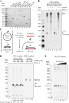
Fig 1
Fig 1. Factors affecting R-loop formation in vitro.
A. Purified, recombinant proteins used in the present study visualized by Stain Free SDS-PAGE (Bio-Rad). B. In vitro transcription from LSP with POLRMT (20 nM), TFAM (200 nM) and TFB2M (60 nM). R-loops were formed and detected as described in panel C. TEFM (40 nM) was added to the indicated reactions. Products formed are labeled as followed: PT: transcripts prematurely terminated at CSBII; RC: longer transcripts formed by rolling circle transcription; and R-loops: transcripts unaffected by RNase A treatment (lane 6). The RNA was labeled by [32P]UTP incorporation. C. Reaction scheme for R-loop formation. A pUC18 plasmid containing an LSP insert, including the CSB region (pUC-LSP, S1 Table) was used. When indicated, the template was treated with topoisomerase I to relax supercoils. In vitro transcription was performed in the presence or absence of TEFM followed by the addition of 300 mM NaCl and RNase A to remove free RNA. D. Effects of mtSSB on in vitro transcription and R-loop formation. Templates used were supercoiled pUC-LSP (lanes 1-6) and as a control, linear pUC-HSP (lanes 7-10, see S1 Table for template sequence). mtSSB concentrations are indicated in nM. HSP RO: Run-off product of HSP transcription; PT: transcripts prematurely terminated at CSBII; and R-loops: transcripts unaffected by RNase A treatment. The ratio of R-loops/CSBII pre-terminated transcripts for each mtSSB concentration is indicated (see Materials and methods). E. R-loop formation was as in 1C, but without RNase A treatment. Increasing RNase H1 concentrations were added (0, 1, 2, 4, 8, 16 and 32 nM in lanes 1-7). PT indicates transcripts prematurely terminated at CSBII.
Fig 2
Fig 2. Replication initiation on supercoiled templates.
A. Reaction scheme for replication initiation reactions combining mitochondrial transcription and replication proteins with supercoiled templates. B. Reactions as described in panel A, with supercoiled pUC19 (lanes 1-3) or pUC-LSP (lanes 4-6) in the presence or absence of POLγ and POLRMT. DNA synthesis products were labeled by [32P]dTTP. Products at the top of the gel are likely due incorporation of radioactive nucleotides at plasmid nicks (“nick translation”), since these products are observed also with POLγ only (Fig 2B, lanes 2 and 5). C. Reactions described in panel A with supercoiled pUC19 (lanes 1-4) or pUC-LSP (lanes 5-8) in the presence of both POLγ and POLRMT. Increasing amounts of mtSSB were added (0, 8, 32, 128 nM in lanes 1-4 and 5-8 respectively). DNA synthesis products were labeled by [32P]dTTP. D. POLγ-dependent DNA synthesis from processed R-loops in reactions as in Fig 2A using the pUC-LSP template. DNA synthesis products were labeled by [32P]dTTP. RNase H1 (0.5, 2 and 8 nM in lanes 1-4, 5-8 and 9-12 respectively) and mtSSB (20 nM in lanes 5-8 or 120 nM in lanes 9-12) were added when indicated. DNA synthesis products formed in presence of RNase H1 and mtSSB are indicated (Putative specific initiations). E. POLγ-dependent DNA synthesis with a pUC19 template. DNA synthesis products were labeled by [32P]dTTP. RNase H1 (2 nM) and mtSSB (120 nM) were added as indicated. F. TEFM abolishes POLγ-dependent DNA synthesis from processed R-loops. Reactions were performed as in panel A and B, with pUC-LSP template in the presence of 120 nM mtSSB, 2 nM RNase H1 and increasing concentrations of TEFM (0, 4, 20 and 100 nM in lanes 1-4 respectively). RNase H1 dependent DNA synthesis products are indicated (Putative specific initiations).
Fig 3
Fig 3. RNase H1 and mtSSB activate R-loop dependent replication initiation.
A. Reaction scheme for R-loop-specific initiation of replication. Replication products were labeled by [32P]dTTP and DNA synthesis was terminated by ddCTP incorporation. KOH treatment was used to remove any ribonucleotide primer residues. Products were separated on denaturing sequencing gels. The same basic reaction was performed in panels B-F, with the indicated modifications. B. Replication initiation in the absence of mtSSB. RNase H1 was added as indicated (lane 2-8, 0.25, 0.5, 1, 2, 4, 8 and 16 nM respectively). C. Replication initiation with constant amounts of RNase H1 (2 nM). mtSSB was added as indicated (lane 2-7, 2.5, 5, 10, 20, 40 and 80 nM respectively). D. Replication initiation with pUC-LSP and mutant derivatives thereof; WT, supercoiled (lane 1), WT relaxed (lane 2), CSBIII mutant (lane 3, see S1 Table), and CSBII mutant (lane 4, see S1 Table). Initiation events sensitive to CSBIII and CSBII, or only to CSBII, are indicated. E. Replication initiation in the absence (lane 1) or presence of increasing amounts of TEFM (5, 10, 20, 40 and 80 nM in lanes 2-6 respectively). F. Replication initiation with WT POLγ and/or the indicated factors. All reactions also contain TFAM, TFB2M and POLγB.
Fig 4
Fig 4. RNase H1 disease mutants are unable to support primer processing and DNA replication initiation.
A. Schematic representation of human RNase H1 with the V142I and A185V mutations indicated. The mitochondrial targeting signal (MTS) is shown in blue, the hybrid binding domain (HBD) in green, the connection domain (CD) in grey and the catalytic domain in red. B. R-loop processing by RNase H1 mutant enzymes. The reactions were performed as in Fig 1E. WT, V142I or A185V RNase H1 enzymes were added at 2, 8 and 32 nM in lane 2-10. Reactions in lanes 11-13 contained both V142I and A185V mutants with total enzyme concentration still at 2, 8 and 32 nM. Lane 1 is a no RNase H1 control. PT indicates transcripts prematurely terminated at CSBII. C. Replication initiation reaction as described in Fig 3, with 40 nM mtSSB and 2 nM RNase H1 WT or mutants as described in panel B.
Fig 5
Fig 5. DNA replication initiation defects in RNase H1 deficient cells.
A. Schematic representation of the control region of mtDNA with the positions of primer extension primers 1 and 2 indicated. B. Primer extension of mtDNA from control and RNase H1 patient cells with primer 1 (corresponding to mtDNA positions 8-29, see panel A). Primer 1 was annealed to mtDNA from control (WT) and RNase H1 patient (mut RH1) cells and extended over the OriH, CSB and LSP regions with Taq DNA polymerase (see method details). Control cell DNA (WT) in lanes 1-2 and RNase H1 patient cell DNA in lanes 3-4. Untreated DNA in lanes 1 and 3, and RNase H2 treated DNA in lanes 2 and 4. The NEB LMW ladder is indicated in black, mtDNA positions in green, mapped 5′-ends in blue and mapped RNA to DNA transition points in red. G, C, A and T sequencing ladders are found on the left-hand side. A schematic representation of the control region with OriH, CSBI-III and LSP is shown on the right-hand side. C. Primer extension of mtDNA from control and RNase H1 patient cells with primer 2 (corresponding to mtDNA positions 16,231-16,251, see panel A). Sample loading and colored indications as in panel B. D. 5′-end sequencing (5′-End-seq) of control cell mtDNA. E. Hydrolytic end sequencing (HydEn-seq) of control cell mtDNA to map 5′-ends with attached ribonucleotides. F. 5′-end sequencing (5′-End-seq) of RNase H1 patient cell mtDNA. G. Hydrolytic end sequencing (HydEn-seq) of RNase H1 patient cell mtDNA to map 5′-ends with attached ribonucleotides.
Fig 6
Fig 6. A simplified model for replication primer formation and replication initiation downstream of LSP.
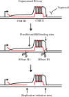
Transcription termination at CSBII leads to the formation of an R-loop, which is stabilized by a G-quadruplex structure formed between the nascent RNA and the G-rich non-template DNA strand. To be used as a primer for mtDNA synthesis, the R-loop must first be processed by RNase H1 to generate 3′-ends, from which POLγ can initiate DNA synthesis. Both R-loop formation and DNA replication initiation are stimulated by mtSSB.

References

    1. Gustafsson CM, Falkenberg M, Larsson NG. Maintenance and Expression of Mammalian Mitochondrial DNA. Annu Rev Biochem. 2016;85:133–60. 10.1146/annurev-biochem-060815-014402 - DOI - PubMed
    1. Falkenberg M, Gaspari M, Rantanen A, Trifunovic A, Larsson NG, Gustafsson CM. Mitochondrial transcription factors B1 and B2 activate transcription of human mtDNA. Nat Genet. 2002;31(3):289–94. 10.1038/ng909 - DOI - PubMed
    1. Minczuk M, He J, Duch AM, Ettema TJ, Chlebowski A, Dzionek K, et al. TEFM (c17orf42) is necessary for transcription of human mtDNA. Nucleic acids research. 2011;39(10):4284–99. 10.1093/nar/gkq1224 - DOI - PMC - PubMed
    1. Agaronyan K, Morozov YI, Anikin M, Temiakov D. Mitochondrial biology. Replication-transcription switch in human mitochondria. Science. 2015;347(6221):548–51. 10.1126/science.aaa0986 - DOI - PMC - PubMed
    1. Posse V, Shahzad S, Falkenberg M, Hallberg BM, Gustafsson CM. TEFM is a potent stimulator of mitochondrial transcription elongation in vitro. Nucleic acids research. 2015;43(5):2615–24. 10.1093/nar/gkv105 - DOI - PMC - PubMed

Publication types