

News about non-secretory exocytosis: mechanisms, properties, and functions
- PMID: 30605539
- PMCID: PMC6821209
- DOI: 10.1093/jmcb/mjy084
News about non-secretory exocytosis: mechanisms, properties, and functions
Abstract
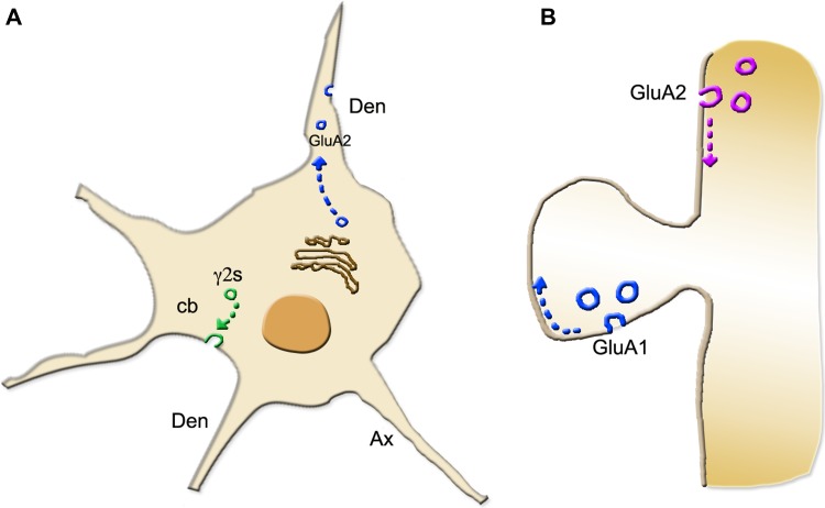
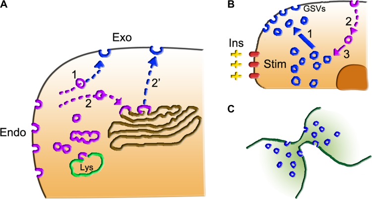
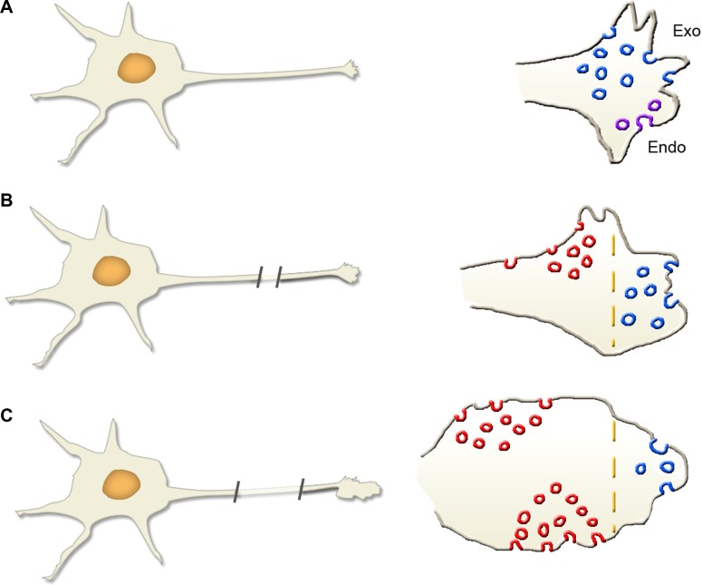
The fusion by exocytosis of many vesicles to the plasma membrane induces the discharge to the extracellular space of their abundant luminal cargoes. Other exocytic vesicles, however, do not contain cargoes, and thus, their fusion is not followed by secretion. Therefore, two distinct processes of exocytosis exist, one secretory and the other non-secretory. The present review deals with the knowledge of non-secretory exocytosis developed during recent years. Among such developments are the dual generation of the exocytic vesicles, initially released either from the trans-Golgi network or by endocytosis; their traffic with activation of receptors, channels, pumps, and transporters; the identification of their tethering and soluble N-ethylmaleimide-sensitive factor attachment protein receptor complexes that govern membrane fusions; the growth of axons and the membrane repair. Examples of potential relevance of these processes for pathology and medicine are also reported. The developments presented here offer interesting chances for future progress in the field.
Keywords: Golgi complex; endocytosis; exocytosis; vesicles.
© The Author(s) (2019). Published by Oxford University Press on behalf of Journal of Molecular Cell Biology, IBCB, SIBS, CAS.
Figures
Similar articles
-
Myosins in the secretory pathway: tethers or transporters?Cell Mol Life Sci. 2008 Sep;65(18):2790-800. doi: 10.1007/s00018-008-8350-5. Cell Mol Life Sci. 2008. PMID: 18726179 Free PMC article. Review.
-
The exocyst complex in polarized exocytosis.Int Rev Cytol. 2004;233:243-65. doi: 10.1016/S0074-7696(04)33006-8. Int Rev Cytol. 2004. PMID: 15037366 Review.
-
The molecular mechanisms of the mammalian exocyst complex in exocytosis.Biochem Soc Trans. 2006 Nov;34(Pt 5):687-90. doi: 10.1042/BST0340687. Biochem Soc Trans. 2006. PMID: 17052175
-
Myosin VI and its binding partner optineurin are involved in secretory vesicle fusion at the plasma membrane.Mol Biol Cell. 2011 Jan 1;22(1):54-65. doi: 10.1091/mbc.E10-06-0553. Epub 2010 Dec 9. Mol Biol Cell. 2011. PMID: 21148290 Free PMC article.
-
How peptide hormone vesicles are transported to the secretion site for exocytosis.Mol Endocrinol. 2008 Dec;22(12):2583-95. doi: 10.1210/me.2008-0209. Epub 2008 Jul 31. Mol Endocrinol. 2008. PMID: 18669645 Free PMC article. Review.
Cited by
-
Polymer Pro-Drug Nanoparticles for Sustained Release of Cytotoxic Drugs Evaluated in Patient-Derived Glioblastoma Cell Lines and In Situ Gelling Formulations.Pharmaceutics. 2021 Feb 3;13(2):208. doi: 10.3390/pharmaceutics13020208. Pharmaceutics. 2021. PMID: 33546301 Free PMC article.
-
Endocytosis and exocytosis protect cells against severe membrane tension variations.Biophys J. 2021 Dec 21;120(24):5521-5529. doi: 10.1016/j.bpj.2021.11.019. Epub 2021 Nov 25. Biophys J. 2021. PMID: 34838532 Free PMC article.
