. 2019 Feb 12;58(6):706-713.
doi: 10.1021/acs.biochem.8b01145. Epub 2019 Jan 15.

Structural and Spectroscopic Characterization of a Product Schiff Base Intermediate in the Reaction of the Quinoprotein Glycine Oxidase, GoxA

Affiliations

Structural and Spectroscopic Characterization of a Product Schiff Base Intermediate in the Reaction of the Quinoprotein Glycine Oxidase, GoxA

Dante Avalos et al. Biochemistry. .

Abstract

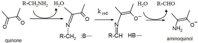
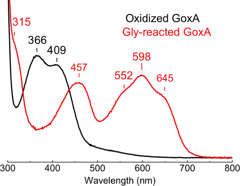
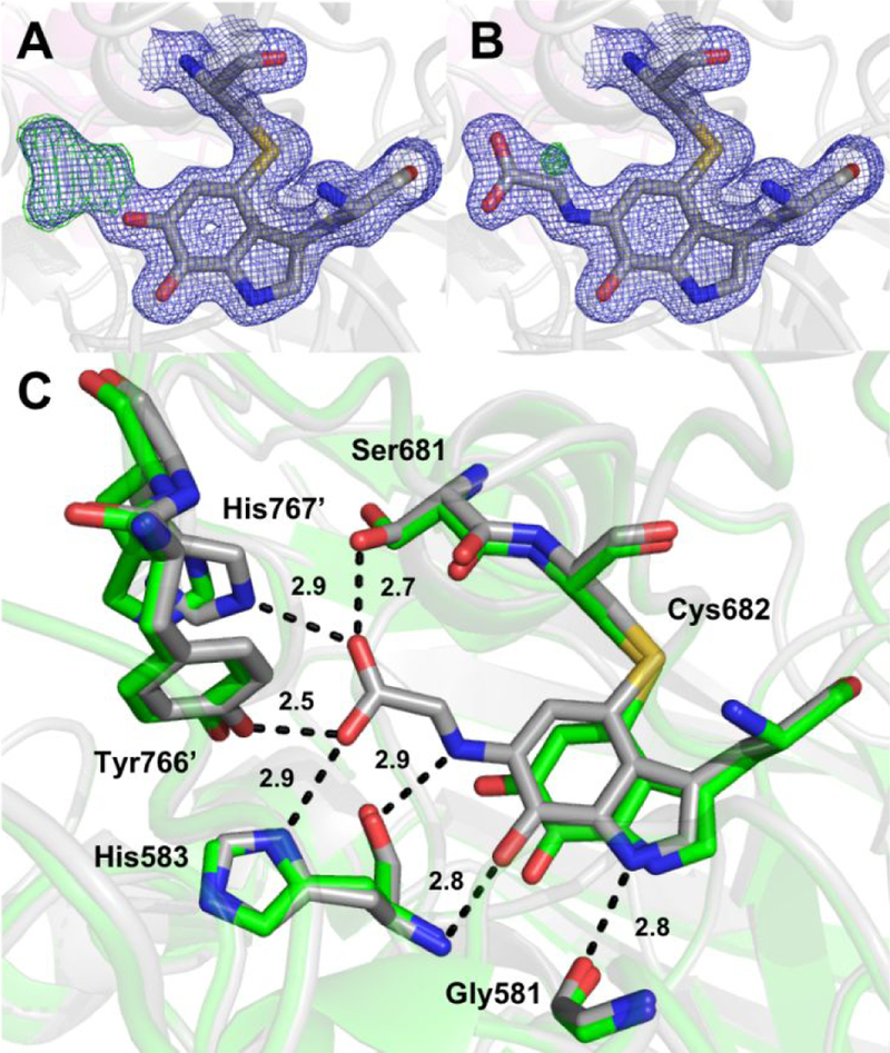
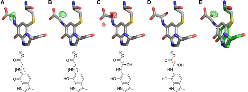
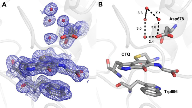
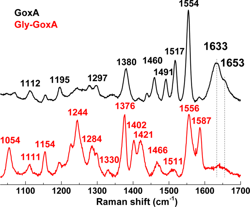
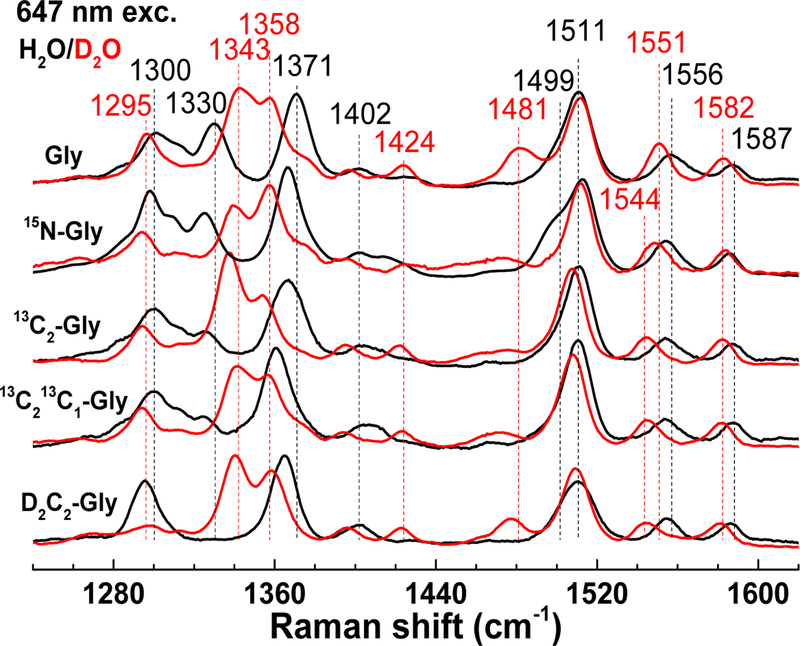
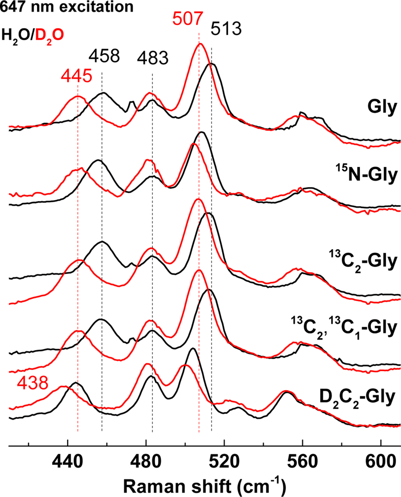
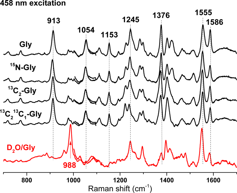
The LodA-like proteins make up a recently identified family of enzymes that rely on a cysteine tryptophylquinone cofactor for catalysis. They differ from other tryptophylquinone enzymes in that they are oxidases rather than dehydrogenases. GoxA is a member of this family that catalyzes the oxidative deamination of glycine. Our previous work with GoxA from Pseudoalteromonas luteoviolacea demonstrated that this protein forms a stable intermediate upon anaerobic incubation with glycine. The spectroscopic properties of this species were unique among those identified for tryptophylquinone enzymes characterized to date. Here we use X-ray crystallography and resonance Raman spectroscopy to identify the GoxA catalytic intermediate as a product Schiff base. Structural work additionally highlights features of the active site pocket that confer substrate specificity, intermediate stabilization, and catalytic activity. The unusual properties of GoxA are discussed within the context of the other tryptophylquinone enzymes.


Figures

Figure 1.
Figure 1.
The reductive half-reaction of tryptophylquinone and topaquinone enzymes. Schiff-base adducts are shown as neutral species as the protonation states are unknown for tryptophylquinone enzymes.
Figure 2.
Figure 2.
Absorption spectra of oxidized GoxA (black) and of the stable intermediate formed after the addition of 5 mM glycine in absence of O2 (Gly-GoxA, red). Samples were in 1-mm Raman capillaries at a concentration of 550 M GoxA in 50 mM potassium phosphate buffer pH 7.5.
Figure 3.
Figure 3.
A GoxA crystal in mother liquor containing 10 mM glycine (A) was subsequently transferred to glycine-free mother liquor and imaged over approximately 5 minutes (B-F)
Figure 4.
Figure 4.
The CTQ site of glycine-soaked GoxA crystals modeled as (A) oxidized CTQ and (B) a product Schiff-base. 2Fo-Fc and Fo-Fc electron density are shown as blue and green mesh contoured to 1.5σ and 5.0σ, respectively. (C) Comparison of the CTQ environment in the product Schiff-base (gray) and oxidized (PDB ID: 6BYW, green) GoxA. Probable hydrogen bonds in the glycine adduct structure are shown as dotted lines with indicated interatomic distances.
Figure 5.
Figure 5.
The glycine adduct modeled as (A) substrate Schiff-base, (B) product Shiff-base, (C) R-hydroxylated intermediate and (D) S-hydroxylated intermediate. (E) The oxidized CTQ and associated water molecule are overlaid on B. Positive and negative Fo-Fc electron density are shown as green and red mesh, respectively, contoured to 4.0 . Square brackets in chemical drawings of (A) and (B) reflect uncertainty of the protonation state of the nitrogen atom.
Figure 6.
Figure 6.
Water structure around the glycine adduct. (A and B) The adduct modeled as the product Schiff-base with relevant residues are shown as sticks and hydrogen bonds as dotted lines with indicated interatomic distances. 2Fo-Fc electron density is shown as blue mesh contoured to 1.0 .
Figure 7.
Figure 7.
RR spectra of oxidized GoxA (black) and Gly-GoxA (red) with 458 nm excitation at room temperature; the final protein concentration was ~550 μM in 50 mM KPi pH 7.5.
Figure 8.
Figure 8.
High-frequency RR spectra of isotopically labeled Gly-GoxA in H2O (black) and D2O (red) with 647 nm excitation at room temperature.
Figure 9.
Figure 9.
Low-frequency RR spectra of isotopically labeled Gly-GoxA in H2O (black) and D2O (red) with 647 nm excitation at room temperature.
Figure 10.
Figure 10.
Combined low- and high-frequency RR spectra of isotopically labeled Gly-GoxA spectra obtained with 458 nm excitation at room temperature.

Similar articles

Cited by

References

    1. McIntire WS, Wemmer DE, Chistoserdov A, and Lidstrom ME (1991) A new cofactor in a prokaryotic enzyme: tryptophan tryptophylquinone as the redox prosthetic group in methylamine dehydrogenase, Science 252, 817–824. - PubMed
    1. Datta S, Mori Y, Takagi K, Kawaguchi K, Chen ZW, Okajima T, Kuroda S, Ikeda T, Kano K, Tanizawa K, and Mathews FS (2001) Structure of a quinohemoprotein amine dehydrogenase with an uncommon redox cofactor and highly unusual crosslinking, Proc. Natl. Acad. Sci. U. S. A 98, 14268–14273. - PMC - PubMed
    1. Satoh A, Kim JK, Miyahara I, Devreese B, Vandenberghe I, Hacisalihoglu A, Okajima T, Kuroda S, Adachi O, Duine JA, Van Beeumen J, Tanizawa K, and Hirotsu K (2002) Crystal structure of quinohemoprotein amine dehydrogenase from Pseudomonas putida. Identification of a novel quinone cofactor encaged by multiple thioether cross-bridges, J. Biol. Chem 277, 2830–2834. - PubMed
    1. Davidson VL (2005) Structure and mechanism of tryptophylquinone enzymes, Bioorg. Chem 33, 159–170. - PubMed
    1. Yukl ET, and Davidson VL (2018) Diversity of structures, catalytic mechanisms and processes of cofactor biosynthesis of tryptophylquinone-bearing enzymes, Arch. Biochem. Biophys 654, 40–46. - PMC - PubMed

Publication types