

The anti-obesity effects of Tongbi-san in a high-fat diet-induced obese mouse model
- PMID: 30606178
- PMCID: PMC6319014
- DOI: 10.1186/s12906-018-2420-5
The anti-obesity effects of Tongbi-san in a high-fat diet-induced obese mouse model
Abstract
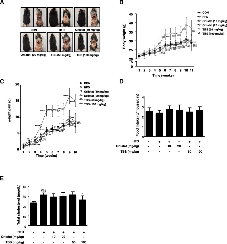
Background: Recently, it has been noted that natural herbal medications may be effective in treating obesity. Tongbi-san (TBS) is a traditional medicine usually used for dysuria (i.e., painful urination), containing three herbs, Cyperus rotundus L., Citrus unshiu Markovich, and Poria cocos. In this study, we aimed to examine whether TBS can inhibit high-fat diet (HFD)-induced adipogenesis in the liver and epididymal adipose tissue of obese mice.
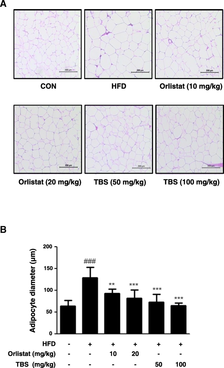
Methods: Male C57BL/6 N mice were fed a normal diet, an HFD, an HFD plus orlistat 10 or 20 mg/kg, or an HFD plus TBS 50 or 100 mg/kg for 11 weeks. Body weight was checked weekly and histological tissue examinations were investigated. An expression of genes involved in adipogenesis was also assessed.
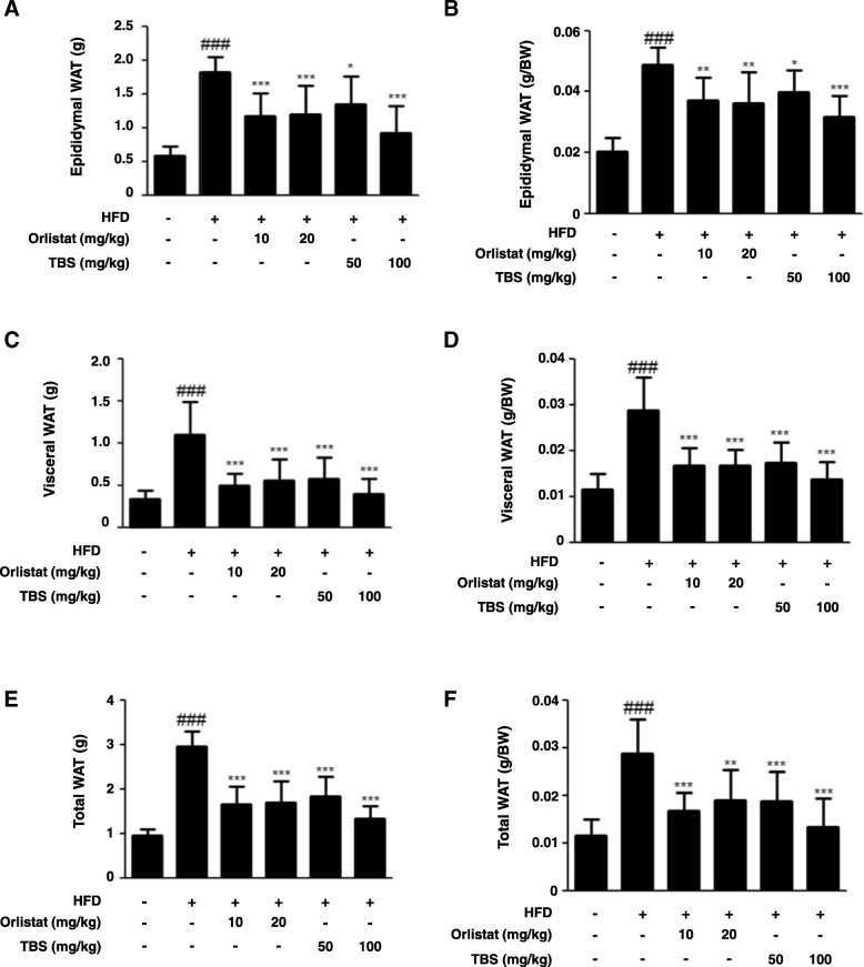
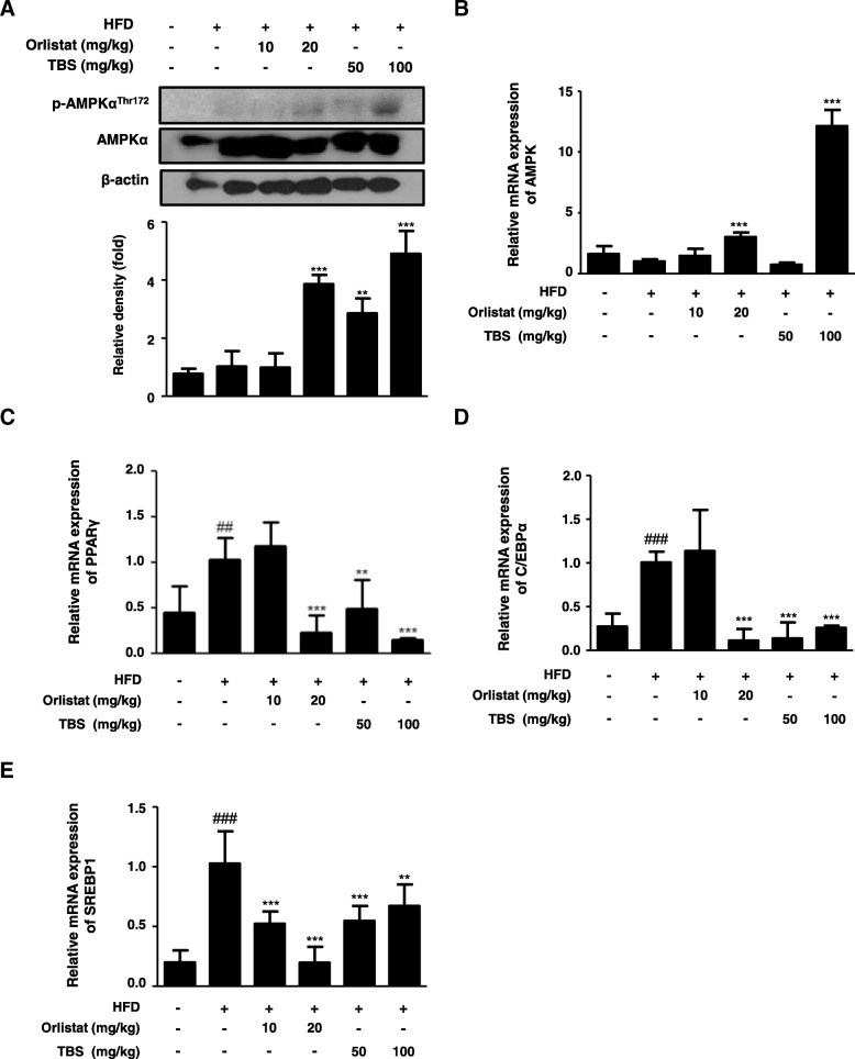
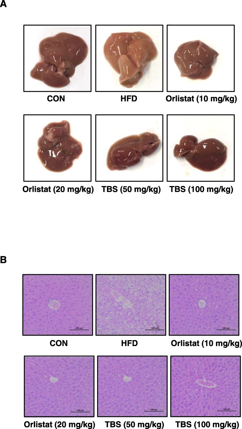
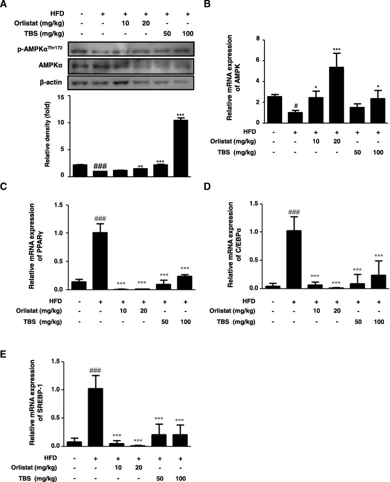
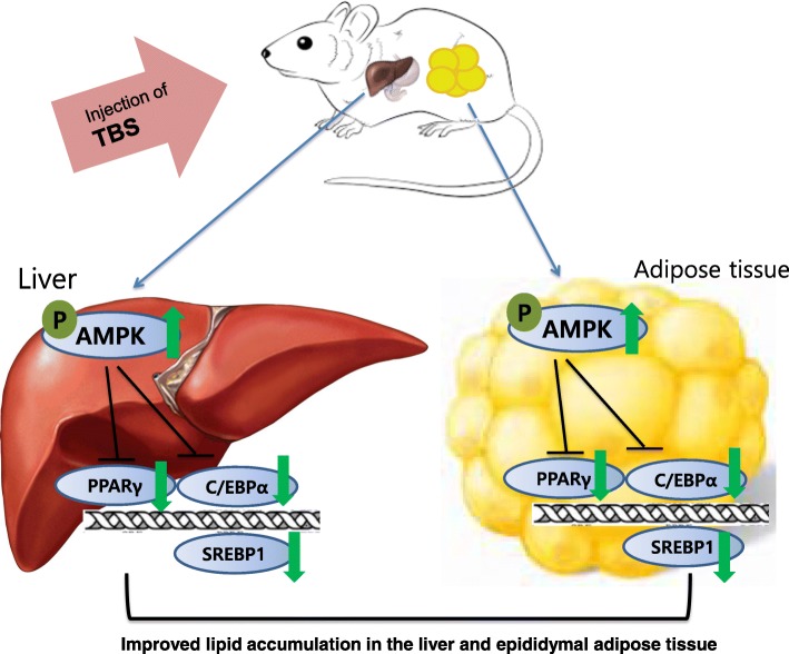
Results: Oral administration of TBS significantly reduced body weight and decreased epididymal and visceral white adipose tissue (WAT) weight. In addition, we found that TBS enhanced the expression of the adenosine monophosphate-activated protein kinase (AMPK) and inhibited the expression of transcription factors, such as CCAAT/enhancer-binding proteins (C/EBPs), sterol regulatory element-binding protein 1 (SREBP1), and peroxisome proliferator-activated receptor γ (PPARγ) in the liver and epididymal WAT as measured by quantitative reverse transcription polymerase chain reaction (qRT-PCR).
Conclusion: These findings demonstrate that the anti-obesity effects of TBS may be linked to the activation of AMPK.
Keywords: AMPK; Adipogenesis; High-fat diet; Obesity; Tongbi-san.
Conflict of interest statement
Ethics approval and consent to participate
All procedures were conducted in accordance with the National Institute of Health guidelines and approved by the Ethical Committee for Animal Care and the Use of Laboratory Animal of Sangji University (reg.no. 2017–12).
Consent for publication
Not applicable.
Competing interests
The authors declare that they have no competing interest.
Publisher’s Note
Springer Nature remains neutral with regard to jurisdictional claims in published maps and institutional affiliations.
Figures
Similar articles
-
HOX-7 suppresses body weight gain and adipogenesis-related gene expression in high-fat-diet-induced obese mice.BMC Complement Altern Med. 2014 Dec 17;14:505. doi: 10.1186/1472-6882-14-505. BMC Complement Altern Med. 2014. PMID: 25515293 Free PMC article.
-
Gynostemma Pentaphyllum Extract Ameliorates High-Fat Diet-Induced Obesity in C57BL/6N Mice by Upregulating SIRT1.Nutrients. 2019 Oct 15;11(10):2475. doi: 10.3390/nu11102475. Nutrients. 2019. PMID: 31618980 Free PMC article.
-
Green tomato extract attenuates high-fat-diet-induced obesity through activation of the AMPK pathway in C57BL/6 mice.J Nutr Biochem. 2013 Jan;24(1):335-42. doi: 10.1016/j.jnutbio.2012.06.018. Epub 2012 Sep 10. J Nutr Biochem. 2013. PMID: 22974972
-
Cyanidin-3-O-galactoside-enriched Aronia melanocarpa extract attenuates weight gain and adipogenic pathways in high-fat diet-induced obese C57BL/6 mice.Nutrients. 2019 May 27;11(5):1190. doi: 10.3390/nu11051190. Nutrients. 2019. PMID: 31137884 Free PMC article.
-
Do the Natural Chemical Compounds Interact with the Same Targets of Current Pharmacotherapy for Weight Management?-A Review.Curr Drug Targets. 2019;20(4):399-411. doi: 10.2174/1389450119666180830125958. Curr Drug Targets. 2019. PMID: 30173643 Review.
Cited by
-
Drynaria rhizome water extract alleviates high‑fat diet‑induced obesity in mice.Mol Med Rep. 2024 Feb;29(2):30. doi: 10.3892/mmr.2023.13153. Epub 2023 Dec 22. Mol Med Rep. 2024. PMID: 38131179 Free PMC article.
-
Response surface methodology based development of an optimized polyherbal formulation and evaluation of its anti-diabetic and anti-obesity potential in high-fat diet-induced obese mice.J Tradit Complement Med. 2023 Jul 16;14(1):70-81. doi: 10.1016/j.jtcme.2023.07.002. eCollection 2024 Jan. J Tradit Complement Med. 2023. PMID: 38223811 Free PMC article.
-
Anti-Obesity Effects of Sargassum thunbergii via Downregulation of Adipogenesis Gene and Upregulation of Thermogenic Genes in High-Fat Diet-Induced Obese Mice.Nutrients. 2020 Oct 29;12(11):3325. doi: 10.3390/nu12113325. Nutrients. 2020. PMID: 33138053 Free PMC article.
-
Impact of Elateriospermum tapos Supplementation on Leptin and Hypothalamic Signaling in Female Offspring of High-Fat Diet-Induced Obese.Adv Pharm Bull. 2025 Apr 4;15(1):186-193. doi: 10.34172/apb.43919. eCollection 2025 Apr. Adv Pharm Bull. 2025. PMID: 40636308 Free PMC article.
-
Magnolia officinalis Rehder & E. Wilson ameliorates white adipogenesis by upregulating AMPK and SIRT1 in vitro and in vivo.Heliyon. 2024 Mar 7;10(6):e27600. doi: 10.1016/j.heliyon.2024.e27600. eCollection 2024 Mar 30. Heliyon. 2024. PMID: 38515723 Free PMC article.
References
-
- Zou T, Wang B, Yang Q, de Avila JM, Zhu MJ, You J, Chen D, Du M. Raspberry promotes brown and beige adipocyte development in mice fed high-fat diet through activation of AMP-activated protein kinase (AMPK) alpha1. J Nutr Biochem. 2018;55:157–164. doi: 10.1016/j.jnutbio.2018.02.005. - DOI - PMC - PubMed
MeSH terms
Substances
Grants and funding
LinkOut - more resources
Full Text Sources
Medical
Research Materials
