Review
. 2019 Jan 3;14(1):1.
doi: 10.1186/s13023-018-0972-6.

Achondroplasia: a comprehensive clinical review

Affiliations
Review

Achondroplasia: a comprehensive clinical review

Richard M Pauli. Orphanet J Rare Dis. .

Abstract

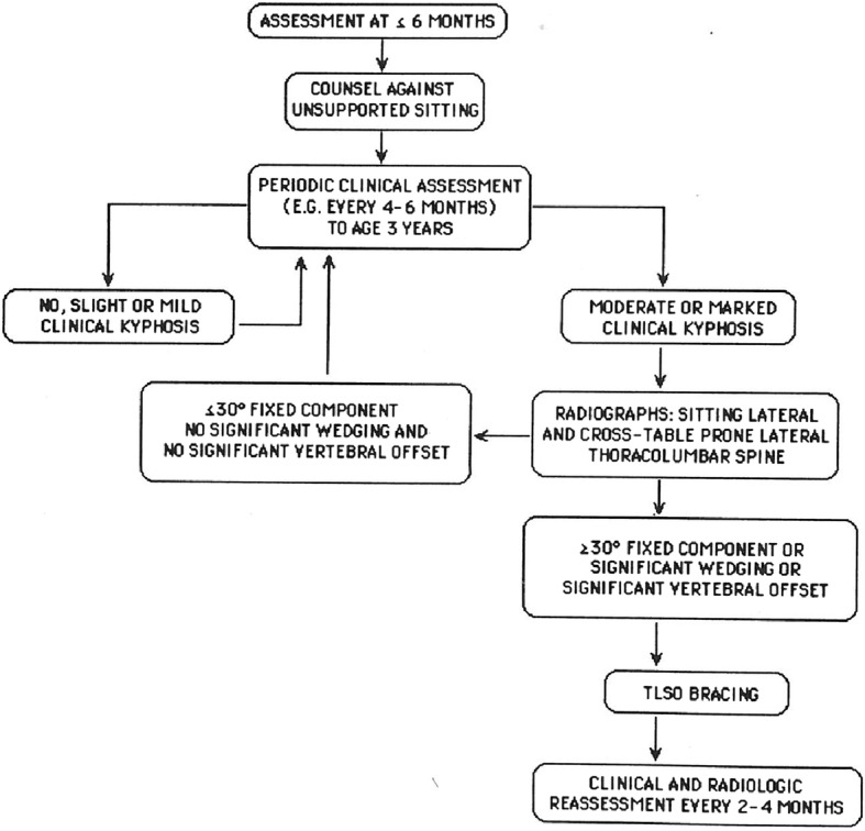
Achondroplasia is the most common of the skeletal dysplasias that result in marked short stature (dwarfism). Although its clinical and radiologic phenotype has been described for more than 50 years, there is still a great deal to be learned about the medical issues that arise secondary to this diagnosis, the manner in which these are best diagnosed and addressed, and whether preventive strategies can ameliorate the problems that can compromise the health and well being of affected individuals. This review provides both an updated discussion of the care needs of those with achondroplasia and an exploration of the limits of evidence that is available regarding care recommendations, controversies that are currently present, and the many areas of ignorance that remain.

Keywords: Achondroplasia; Care guidelines; FGFR3; Natural history; Skeletal dysplasia.


Conflict of interest statement

Ethics approval and consent to participate

No new human data is used in this review and therefore no ethics approval is applicable.

Consent for publication

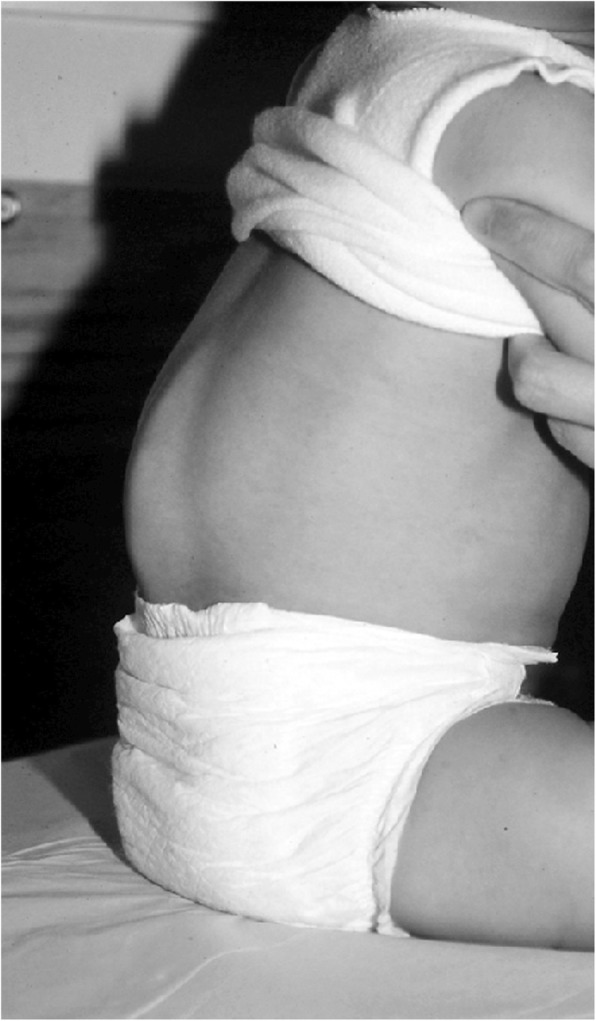
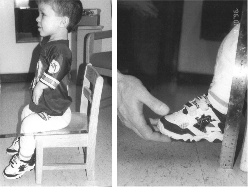
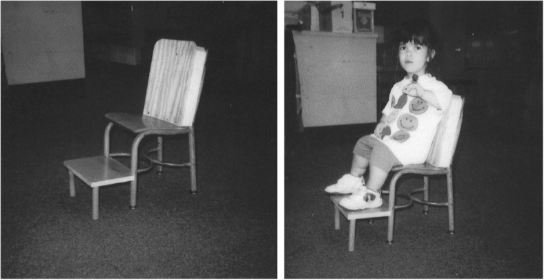
Written informed consent was obtained from individuals or their guardians for publication of all recognizable images. A copy of those written consents is available for review by the Editor-in-Chief of this journal.

Competing interests

The author declares that he has no competing interests.

Publisher’s Note

Springer Nature remains neutral with regard to jurisdictional claims in published maps and institutional affiliations.

Figures

Fig. 1
Fig. 1
Simplified diagram of the FGFR3 protein, including three immunoglobulin-like domains (Ig), a transmembrane domain (TM), and the split tyrosine kinase (TK) region
Fig. 2
Fig. 2
The body phenotype is shown in individuals of different ages: Left to right – infancy, early childhood, childhood and adulthood. In all, note the rhizomelic shortening of the limbs, which are disproportionately short compared with the trunk. In the infant and young child macrocephaly is evident
Fig. 3
Fig. 3
Six portraits of children with achondroplasia. The variability of craniofacial features is evident. Lower left and lower center photographs originally published in Pauli RM (1995) Osteochondrodysplasias with mild clinical manifestations: A guide for endocrinologists and others. Growth Genet Horm 11:1–5
Fig. 4
Fig. 4
Anteroposterior radiograph of the pelvis and femora in an infant with achondroplasia. Characteristic findings include a squared-off pelvis, horizontal acetabula, very narrow sacrosciatic notch, characteristic proximal femoral radiolucency, and short and robust femora
Fig. 5
Fig. 5
Arm radiograph in a newborn with achondroplasia. Although there are generalized metaphyseal abnormalities and shortening of all of the long bones, characteristics here are not as diagnostically helpful as those shown in Fig. 4
Fig. 6
Fig. 6
Hands in achondroplasia, well illustrating brachydactyly and (here, asymmetric) trident configuration – excess separation between the third and fourth fingers. Originally published in Pauli RM (2010) Achondroplasia. In Cassidy SB, Allanson JE: Management of Genetic Syndromes, 3rd ed. Wiley-Blackwell
Fig. 7
Fig. 7
General body habitus present in hypochondroplasia. Cursory examination could easily miss the presence of a bone growth disorder in such a child. Originally published in Pauli RM (1995) Osteochondrodysplasias with mild clinical manifestations: A guide for endocrinologists and others. Growth Genet Horm 11:1–5
Fig. 8
Fig. 8
Clinical phenotype of thanatophoric dysplasia, type I. All features are far more severe than those seen in babies with achondroplasia (Fig. 2)
Fig. 9
Fig. 9
Anteroposterior radiograph of the pelvis and femora in thanatophoric dysplasia, type I. Here, too, qualitatively most of the abnormal characteristics are similar to those seen in achondroplasia, but quantitatively all of them are much more severe. Note the so-called telephone receiver femora
Fig. 10
Fig. 10
Siblings. On the left is an infant with typical, heterozygous achondroplasia. On the right is his older sister who had homozygous achondroplasia. Note the far greater limb foreshortening and much smaller stature in the latter
Fig. 11
Fig. 11
Gross pathologic features from the craniocervical junction of the spinal cord in an infant with achondroplasia who died suddenly and unexpectedly. There is gross indentation of the cord as well as cystic lesions secondary to hypoxic damage. Originally published in Pauli RM et al. (1984) Apnea and sudden unexpected death in infants with achondroplasia. J Pediatr 104:342–348 [113]
Fig. 12
Fig. 12
Histologic findings from the cervicomedullary junction in the infant described in the text. Left shows severe pyknosis secondary to hypoxic damage, compared with, right, a normal control of comparable age. Originally published in Pauli RM et al. (1984) Apnea and sudden unexpected death in infants with achondroplasia. J Pediatr 104:342–348 [113]
Fig. 13
Fig. 13
Computerized tomography in five infants with achondroplasia, demonstrating the variability of conformation of the foramen magnum
Fig. 14
Fig. 14
Sagittal views of magnetic resonance imaging of the cervical cord in four infants with achondroplasia. a There is obliteration of the posterior subarachnoid fluid layer; b In addition to obliteration of the fluid layer posteriorly, there is “nicking” of the posterior cord; c Narrowing of the cord is evident secondary to “waisting” – that is, there is some indentation anteriorly, as well; d An obvious T2 signal abnormality is present
Fig. 15
Fig. 15
Diagnostic specific linear growth charts for females (left) and males (right) with achondroplasia. Comparable curves for average statured individuals are shaded. Reproduced with permission from Greenwood Genetics Center (1988) Growth References from Conception to Adulthood. Clinton, SC: Jacobs [156]
Fig. 16
Fig. 16
Diagnostic specific weight charts for children 0–36 months (left) and from 2 to 16 years (right). Reproduced with permission from Hoover-Fong JE et al., (2007) Weight for age charts for children with achondroplasia. Am J Med Genet A 143A:2227–2235 [173]
Fig. 17
Fig. 17
Weight by height charts for children with achondroplasia up to 104 cm in height. a Top is for males and b lower is for females. Reproduced with permission from Hunter et al. (1996) Standard weight for height curves in achondroplasia. Am J Med Genet 62:255–261 [174]
Fig. 18
Fig. 18
Weight by height charts for individuals with achondroplasia greater than 104 cm in height. a Top is for males and b lower is for females. Reproduced with permission from Hunter et al. (1996) Standard weight for height curves in achondroplasia. Am J Med Genet 62:255–261 [174]
Fig. 19
Fig. 19
Diagnosis-specific body mass index standards for children with achondroplasia. a Top is for males and b bottom for females. Reproduced with permission from Hoover-Fong JE et al. (2008) Age-appropriate body mass index in children with achondroplasia: interpretation in relation to indexes of height. Am J Clin Nutr 88:364–371 [176]
Fig. 20
Fig. 20
Limited elbow extension in a young child with achondroplasia
Fig. 21
Fig. 21
Marked rhizomelia in a child with achondroplasia
Fig. 22
Fig. 22
Position – remarkably, a comfortable one – illustrating the large joint hypermobility that is present in younger children with achondroplasia
Fig. 23
Fig. 23
Snowplowing. As described in the text, movement is effected by pushing with the feet, sliding the head forward
Fig. 24
Fig. 24
Reverse snowplowing. Here pushing with the heels propels the child who is also supported by the back of the head. Originally published in Fowler ES et al. (1997) Biophysical bases for delayed and aberrant motor development in young children with achondroplasia. J Dev Behav Pediatr 18:143–150 [184]
Fig. 25
Fig. 25
Preorthograde motor movement strategies for infants with achondroplasia. Originally published in Fowler ES et al. (1997) Biophysical bases for delayed and aberrant motor development in young children with achondroplasia. J Dev Behav Pediatr 18:143–150 [184]
Fig. 26
Fig. 26
Demonstration of wrist hypermobility in a school-aged child with achondroplasia. a Positive thumb sign (with wrist flexion the thumb touches the wrist); b positive envelope sign (with wrist flexion all of the fingers can touch the wrist); and c positive 5th finger sign (with wrist extension the 5th finger can be brought parallel to the wrist)
Fig. 27
Fig. 27
Four-finger grasp. Originally published in Fowler ES et al. (1997) Biophysical bases for delayed and aberrant motor development in young children with achondroplasia. J Dev Behav Pediatr 18:143–150 [184]
Fig. 28
Fig. 28
Two-finger grasp taking advantage of the trident configuration gap between the third and fourth fingers. Originally published in Fowler ES et al. (1997) Biophysical bases for delayed and aberrant motor development in young children with achondroplasia. J Dev Behav Pediatr 18:143–150 [184]
Fig. 29
Fig. 29
A developmental screening tool developed by Ireland et al. It is currently the best alternative for developmental screening of children with achondroplasia. Reproduced with permission from Ireland PJ et al., (2012) Development in children with achondroplasia: a prospective clinical cohort study. Dev Med Child Neurol 54:532–537 [9]
Fig. 30
Fig. 30
Head circumference reference standards for females (left) and males (right) with achondroplasia. Comparable measurements for average stature individuals are shaded. Reproduced with permission from Greenwood Genetics Center (1988) Growth References from Conception to Adulthood. Clinton, SC: Jacobs [156]
Fig. 31
Fig. 31
Sequential head circumference measurements in a boy with achondroplasia. Transient acceleration of head growth occurred at around 4–5 years of age (accompanied by non-specific symptoms including occasional emesis). Neuroimaging at that age did demonstrate increased ventricular size compared with imaging completed in the first year of life. There was subsequent equilibration of head growth without intervention. Now an adult, the individual is of normal intelligence and without any indicators of any harmful effects of this transient acceleration and re-equilibration
Fig. 32
Fig. 32
Typical superficial venous prominence in an infant with achondroplasia. This arises from increased flow through emissary veins secondary to increased resistance to flow at the jugular foramina
Fig. 33
Fig. 33
Typical flexible kyphosis seen in infants and young children with achondroplasia
Fig. 34
Fig. 34
Lateral radiograph of the spine of an infant with achondroplasia. While there is obvious kyphosis, there are no secondary changes of the vertebral bodies at the apex of the curve
Fig. 35
Fig. 35
Severe, fixed angular kyphosis of the type that can be prevented by appropriate counseling and intervention in early childhood. Originally published in Pauli RM et al. (1997) Prevention of fixed, angular kyphosis in achondroplasia. J Pediatr Orthop 17:726–733 [64]
Fig. 36
Fig. 36
Cross table supine over-a-bolster lateral radiograph shows a mild irreversible kyphotic curve and mild loss of anterior substance of two vertebrae. This view or, alternatively, a cross table prone lateral radiograph, can be used to assess the irreversible component of kyphotic curvature
Fig. 37
Fig. 37
Suggested algorithm for the assessment and management of kyphosis in infants and young children with achondroplasia. Originally published in Pauli RM et al. (1997) Prevention of fixed, angular kyphosis in achondroplasia. J Pediatr Orthop 17:726–733 [64]
Fig. 38
Fig. 38
Lateral radiograph of the lumbar and sacral spine. It shows the horizontal sacrum and marked hyperlordosis often seen in those with achondroplasia
Fig. 39
Fig. 39
Methods that can be used to monitor progression of varus deformity without repeated radiologic studies (which, however, are needed if the bowing is sufficiently severe that intervention is being considered). ac Measurements between the knees, mid-tibiae and medial malleoli with the legs straight and at rest and the feet together; d Measurement of maximal varus angle by placing the goniometer at the approximate apex of the tibial bow; e Measuring of thigh-foot angle, that is, the angle made by the longitudinal axis of the thigh and the longitudinal axis of the foot when the foot is in its neutral position; this assesses the presence of and severity of internal tibial torsion; f Rough assessment of whether the three joints of the leg are in plumb
Fig. 40
Fig. 40
Two of the common occlusional abnormalities seen in children with achondroplasia. On the left note the narrow, V-shaped anterior palate and the resulting “palisading” of the upper incisors. The right photograph shows a severe anterior open bite
Fig. 41
Fig. 41
On the left – Demonstration of the difficulty a typical child with achondroplasia has when sitting in a school chair. Note that there is neither back nor foot support. Method of measuring for back support is also shown. On the right – measurement that can be made to guide modification to allow foot support
Fig. 42
Fig. 42
Left – Example of modifications made in a school chair to allow for back and foot support. Right – Resultant supported, comfortable sitting for a child with achondroplasia
Fig. 43
Fig. 43
Example of a foldable ‘bottom-wiper’ that can be carried in a pocket or purse and used for personal hygiene by wrapping toilet paper around its end, and swishing it in the toilet to dispose of the paper after its use

References

    1. Trotter TL, Hall JG. Health supervision for children with achondroplasia. Pediatrics. 2005;116:771–781. doi: 10.1542/peds.2005-1440. - DOI - PubMed
    1. Wright MJ, Irving MD. Clinical management of achondroplasia. Arch Dis Child. 2012;97:129–134. doi: 10.1136/adc.2010.189092. - DOI - PubMed
    1. Ireland PJ, Pacey V, Zankl A, Edward P, Johnston LM, Savarirayan R. Optimal management of complications associated with achondroplasia. Appl Clin Genet. 2014;7:117–125. doi: 10.2147/TACG.S51485. - DOI - PMC - PubMed
    1. Pauli RM, Botto LD. Achondroplasia. In: Cassidy SB, Battaglia A, Carey J, Viskochil DH, editors. Management of Genetic Syndromes. 4. New York: Wiley; 2018.
    1. Horton WA, Hall JG, Hecht JT. Achondroplasia. Lancet. 2007;370:162–172. doi: 10.1016/S0140-6736(07)61090-3. - DOI - PubMed

Publication types

MeSH terms

Substances

LinkOut - more resources