Clinical Trial
. 2019 Mar;47(3):283-294.
doi: 10.1124/dmd.118.085670. Epub 2019 Jan 3.

Continuum of Host-Gut Microbial Co-metabolism: Host CYP3A4/3A7 are Responsible for Tertiary Oxidations of Deoxycholate Species

Affiliations
Clinical Trial

Continuum of Host-Gut Microbial Co-metabolism: Host CYP3A4/3A7 are Responsible for Tertiary Oxidations of Deoxycholate Species

Jian Zhang et al. Drug Metab Dispos. 2019 Mar.

Abstract

The gut microbiota modifies endogenous primary bile acids (BAs) to produce exogenous secondary BAs, which may be further metabolized by cytochrome P450 enzymes (P450s). Our primary aim was to examine how the host adapts to the stress of microbe-derived secondary BAs by P450-mediated oxidative modifications on the steroid nucleus. Five unconjugated tri-hydroxyl BAs that were structurally and/or biologically associated with deoxycholate (DCA) were determined in human biologic samples by liquid chromatography-tandem mass spectrometry in combination with enzyme-digestion techniques. They were identified as DCA-19-ol, DCA-6β-ol, DCA-5β-ol, DCA-6α-ol, DCA-1β-ol, and DCA-4β-ol based on matching in-laboratory synthesized standards. Metabolic inhibition assays in human liver microsomes and recombinant P450 assays revealed that CYP3A4 and CYP3A7 were responsible for the regioselective oxidations of both DCA and its conjugated forms, glycodeoxycholate (GDCA) and taurodeoxycholate (TDCA). The modification of secondary BAs to tertiary BAs defines a host liver (primary BAs)-gut microbiota (secondary BAs)-host liver (tertiary BAs) axis. The regioselective oxidations of DCA, GDCA, and TDCA by CYP3A4 and CYP3A7 may help eliminate host-toxic DCA species. The 19- and 4β-hydroxylation of DCA species demonstrated outstanding CYP3A7 selectivity and may be useful as indicators of CYP3A7 activity.


Figures

None
Graphical abstract
Fig. 1.
Fig. 1.
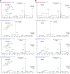
Unconjugated tri-hydroxyl BAs (TBA05, TBA09, TBA10, TBA13, and TBA18) are biologically associated with DCA. The extracted ion chromatograms of the mixed BA standards, whose abbreviations and structures are summarized in Supplemental Table S1 (A). TBA05, TBA09, TBA10, TBA13, and TBA18 were detected in the 0- to 2-hour postprandial urine sample from a representative healthy subject (B). TBA09, TBA10, TBA13, and TBA18 were detected in the 1-hour postprandial serum sample from the same subject (C). Four aliquots of the same urine and serum samples were prepared without enzymes (T1), with choloylglycine hydrolase (T2), with sulfatase and β-glucuronidase (T3), and with all three enzymes (T4). The biologic associations of TBA05, TBA09, TBA10, and TBA13 with DCA were highlighted by hierarchical cluster analysis (D) of the Pearson correlation coefficients between the urinary total unconjugated BAs profile in the test population of 58 healthy volunteers (36 men and 22 women).
Fig. 2.
Fig. 2.
High-definition MS/MS spectra of the oxidized DCA metabolites captured from the synthesized standards (a) and the metabolites detected in vivo or in vitro (b). All standards were synthesized in the laboratory except 3-dehydroDCA was obtained from TRC (cat. no. O856870). The data of endogenous DCA-6β-ol, DCA-5β-ol, DCA-6α-ol, DCA-1β-ol, and DCA-4β-ol were captured from a representative digested urine sample. The in vitro data of DCA-19-ol and 3-dehydroDCA were captured from the DCA incubation samples in human liver microsomes.
Fig. 2.
Fig. 2.
High-definition MS/MS spectra of the oxidized DCA metabolites captured from the synthesized standards (a) and the metabolites detected in vivo or in vitro (b). All standards were synthesized in the laboratory except 3-dehydroDCA was obtained from TRC (cat. no. O856870). The data of endogenous DCA-6β-ol, DCA-5β-ol, DCA-6α-ol, DCA-1β-ol, and DCA-4β-ol were captured from a representative digested urine sample. The in vitro data of DCA-19-ol and 3-dehydroDCA were captured from the DCA incubation samples in human liver microsomes.
Fig. 3.
Fig. 3.
In vitro regioselective oxidation of DCA in human liver microsomes. TBA04, TBA05, TBA09, TBA10, TBA13, TBA18 (m/z 407 > 407), and 3-dehydroDCA (m/z 389 > 389) were detected after incubation of DCA (m/z 391 > 391) in human liver microsomes (A). The retention data of DCA-19-ol, DCA-6β-ol, DCA-5β-ol, DCA-6α-ol, DCA-1β-ol, DCA-4β-ol, and 3-dehydroDCA corresponded well with the metabolites of DCA (B). The identified oxidation sites cluster on the same plane around the 5β-hydrogen in the three-dimensional structure of DCA (C). The metabolite formation kinetics after incubation of 50 µM DCA (n = 3, data shown as mean ± S.D.) in human liver microsomes and human intestinal microsomes for 240 minutes at a protein level of 0.5 mg/ml (D). The inhibition of metabolite formation after incubation of 50 µM DCA in human liver microsomes for 60 minutes by time-dependent inhibitors (n = 3, data shown as mean ± S.D., student t test compared with control: *P < 0.05; **P < 0.01; ***P < 0.001), including verapamil (25 µM), paroxetine (10 µM), ticlopidine (1 µM), tienilic acid (10 µM), phenelzine (1 µM), thio-TEPA (5 µM), and furafylline (1 µM) (E). The inhibition of metabolite formation after incubation of 50 µM DCA in human liver microsomes for 60 minutes by selective inhibitors, including ketoconazole (0.5 µM), fluconazole (10 µM), DEDC (20 µM), quinidine (1 µM), nootkatone (1 µM), sulfaphenazole (1 µM), quercetin (1 µM), montelukast (0.5 µM), sertraline (10 µM), 2-PCPA (0.2 µM), and α-NF (0.1 µM) (F).
Fig. 4.
Fig. 4.
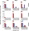
CYP3A4 and CYP3A7 are responsible for the oxidation of DCA, GDCA, and TDCA. The metabolite formation after incubations of 50 µM DCA (A), GDCA (B), and TDCA (C) for 60 minutes in a panel of 18 rCYPs (CYP1A2, 1B1, 2A6, 2B6, 2C8, 2C9*1, 2C18, 2C19, 2D6*1, 3A4, 3A5, 3A7, 2E1, 2J2, 4A11, 4F2, 4F3B, and 4F12; 50 pmol protein/ml) were compared with the data of human liver microsomes (0.5 mg protein/ml) acquired in parallel. The oxidized metabolites of GDCA and TDCA were detected after being digested by choloylglycine hydrolase. Data were shown as mean ± S.D. (n = 3).
Fig. 5.
Fig. 5.
Regioselectivity of CYP3A4, CYP3A5, and CYP3A7 for the oxidation of DCA, GDCA, and TDCA. Metabolite formation after incubations of 50 µM DCA, GDCA, and TDCA for 60 minutes in rCYP3A4, 3A5, and 3A7 (50 pmol protein/ml). The oxidized metabolites of GDCA and TDCA were detected after being digested by choloylglycine hydrolase. Data were shown as mean ± S.D. (n = 3).
Fig. 6.
Fig. 6.
Host-gut microbial cometabolism kinetics of primary, secondary, and tertiary BAs in healthy adults. The time-dependent serum concentrations and their 0- to 2-hour urinary levels (mean ± S.E.M.) were shown for the downstream metabolites of CA (A) and CDCA (B) during the 2-hour postprandial period after a high-fat diet in 13 healthy volunteers. The conjugation pattern (C) was determined by the enzyme digestion techniques, in which the free unconjugated form was detected by T1; the glycine/taurine amidated forms were detected by T2-T1; the glucuronidated/sulfated forms were detected by T3-T1; and the “double-conjugates” linked with both glycine/taurine and glucuronide/sulfate were detected by T4-(T3-T1)-(T2-T1). The apparent renal clearances (D) were calculated based on the total urinary excretion data and the total serum AUC data during the 2-hour postprandial period.

References

    1. Amuro Y, Yamade W, Yamamoto T, Kudo K, Fujikura M, Maebo A, Hada T, Higashino K. (1986) Isocholic acid formation from 7 alpha,12 alpha-dihydroxy-3-keto-5 beta-cholanoic acid with human liver enzyme. Biochim Biophys Acta 879:362–368. - PubMed
    1. Araya Z, Wikvall K. (1999) 6alpha-hydroxylation of taurochenodeoxycholic acid and lithocholic acid by CYP3A4 in human liver microsomes. Biochim Biophys Acta 1438:47–54. - PubMed
    1. Bácsi K, Kósa JP, Borgulya G, Balla B, Lazáry A, Nagy Z, Horváth C, Speer G, Lakatos P. (2007) CYP3A7*1C polymorphism, serum dehydroepiandrosterone sulfate level, and bone mineral density in postmenopausal women. Calcif Tissue Int 80:154–159. - PubMed
    1. Beaumont RN, Warrington NM, Cavadino A, Tyrrell J, Nodzenski M, Horikoshi M, Geller F, Myhre R, Richmond RC, Paternoster L, et al. Early Growth Genetics (EGG) Consortium (2018) Genome-wide association study of offspring birth weight in 86 577 women identifies five novel loci and highlights maternal genetic effects that are independent of fetal genetics. Hum Mol Genet 27:742–756. - PMC - PubMed
    1. Bodin K, Lindbom U, Diczfalusy U. (2005) Novel pathways of bile acid metabolism involving CYP3A4. Biochim Biophys Acta 1687:84–93. - PubMed

Publication types

LinkOut - more resources