. 2019 Feb 27;39(9):1621-1630.
doi: 10.1523/JNEUROSCI.1497-18.2018. Epub 2019 Jan 3.

A Distinct Visual Pathway Mediates High-Intensity Light Adaptation of the Circadian Clock in Drosophila

Affiliations

A Distinct Visual Pathway Mediates High-Intensity Light Adaptation of the Circadian Clock in Drosophila

Matthias Schlichting et al. J Neurosci. .

Abstract

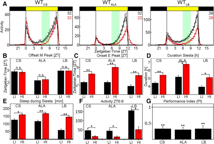
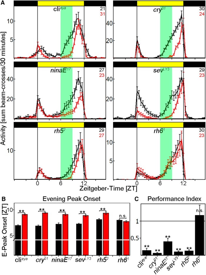
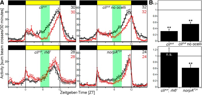
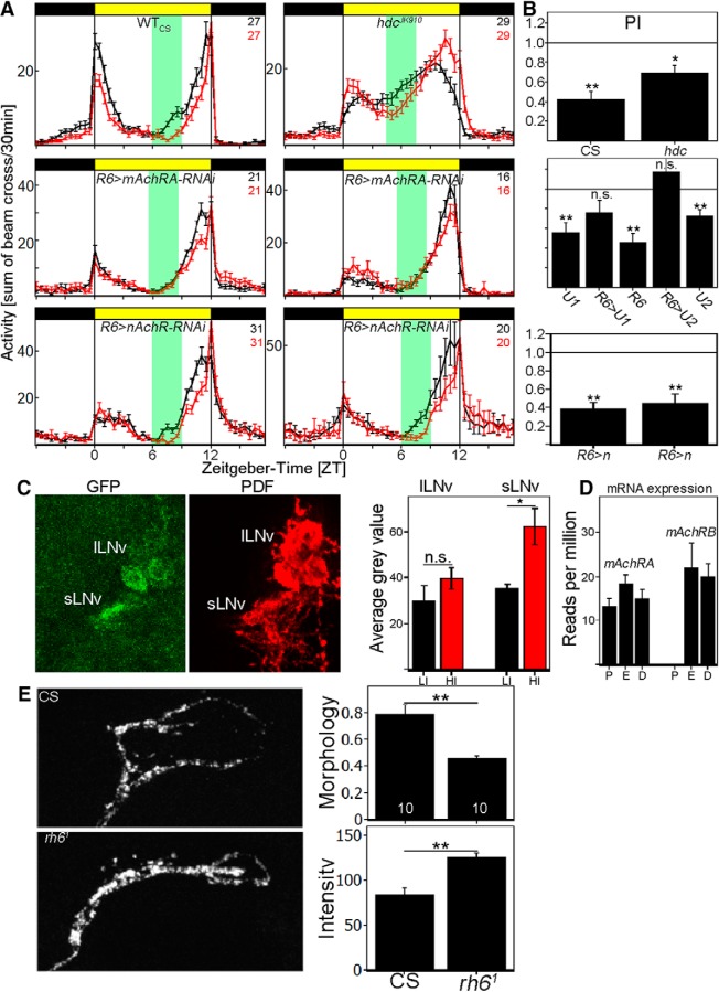
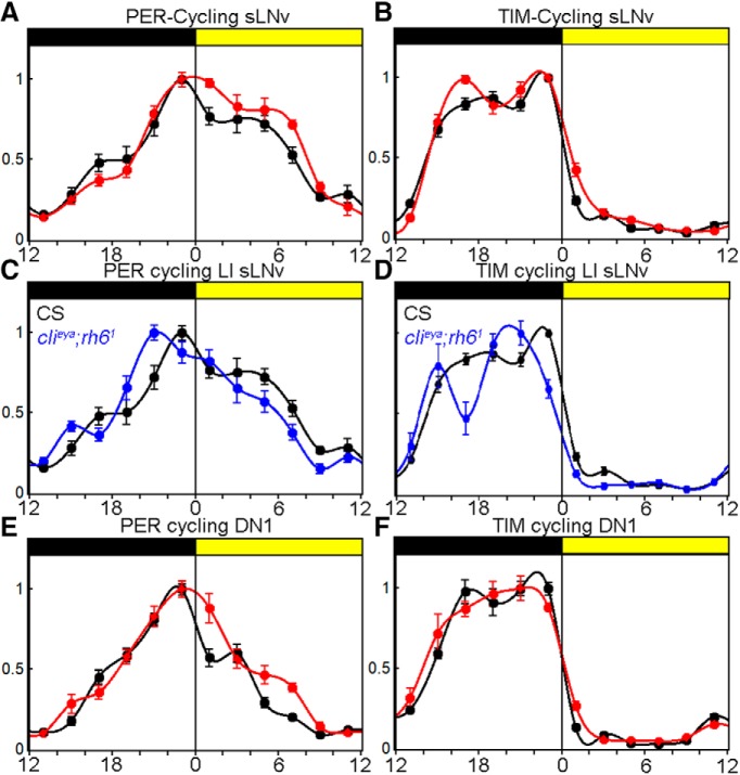
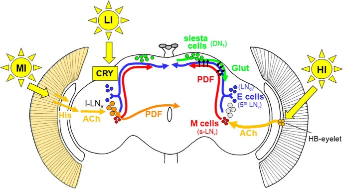
To provide organisms with a fitness advantage, circadian clocks have to react appropriately to changes in their environment. High-intensity (HI) light plays an essential role in the adaptation to hot summer days, which especially endanger insects of desiccation or prey visibility. Here, we show that solely increasing light intensity leads to an increased midday siesta in Drosophila behavior. Interestingly, this change is independent of the fly's circadian photoreceptor cryptochrome and is solely caused by a small visual organ, the Hofbauer-Buchner eyelets. Using receptor knock-downs, immunostaining, and recently developed calcium tools, we show that the eyelets activate key core clock neurons, namely the s-LNvs, at HI. This activation delays the decrease of PERIOD (PER) in the middle of the day and propagates to downstream target clock neurons that prolong the siesta. We show a new pathway for integrating light-intensity information into the clock network, suggesting new network properties and surprising parallels between Drosophila and the mammalian system.SIGNIFICANCE STATEMENT The ability of animals to adapt to their ever-changing environment plays an important role in their fitness. A key player in this adaptation is the circadian clock. For animals to predict the changes of day and night, they must constantly monitor, detect and incorporate changes in the environment. The appropriate incorporation and reaction to high-intensity (HI) light is of special importance for insects because they might suffer from desiccation during hot summer days. We show here that different photoreceptors have specialized functions to integrate low-intensity, medium-intensity, or HI light into the circadian system in Drosophila These results show surprising parallels to mammalian mechanisms, which also use different photoreceptor subtypes to respond to different light intensities.

Keywords: Drosophila; circadian clock; photoreceptor.


Figures

Figure 1.
Figure 1.
Behavior of WT-flies at LI (black line) and HI (red line). A, Average activity profile of WTCS (left), WTALA (middle), and WTLB (right) at LI (10 lux, black line) and HI (10,000 lux, red line) ± SEM recorded in the Regensburg system. All flies show a bimodal activity pattern with reduced activity during the siesta. B, Quantification of M-peak offset at LI (black) and HI (red) in ZT ± SEM. All strains show a M-peak offset ∼2–3 h after lights on, but do not change the onset upon HI (WTCS: p = 0.9379, WTALA: p = 0.1859, WTLB: p = 0.3862). C, Quantification of E-peak onset at LI (black) and HI (red) in ZT ± SEM. All flies significantly delay the E-peak onset by ∼1 h upon HI simulation (p < 0.0001 for all genotypes). D, Duration of siesta in hours ± SEM. All flies show a significantly longer siesta at HI compared with LI (WTCS: p = 0.0001, WTALA: p = 0.0015, WTLB: p = 0.0012). E, Total sleep amount at LI (black) and HI (red) between ZT6 and ZT9 in minutes ± SEM. All flies significantly increase their sleep amounts at HI (WTCS: p = 0.0014, WTALA: p < 0.0001, WTLB: p < 0.0001). F, Sum of activity indicated in the green area in A. All flies significantly reduce their activity level at HI (WTCS: p = 0.0091, WTALA: p = 0.0114, WTLB: p = 0.0006). G, PI calculated by the sum of activity between ZT6 and ZT9 at HI divided by the sum of activity between ZT6 and ZT9 at LI. All genotypes show a PI significantly <1, which we interpret as a delayed E-peak onset (p < 0.0001 for all genotypes). *p < 0.05, **p < 0.001.
Figure 2.
Figure 2.
Behavior of photoreceptor mutants at LI and HI. A, Average activity profiles of flies exposed to LI (black line) and HI (red line) ± SEM using the Regensburg system. Flies deficient of either compound eyes (clieya) or CRY (cry01) behave similar to WTCS, whereas the loss of rhodopsin 6 renders the flies insensitive to light-intensity changes. Consistent with the clieya data, flies affecting the compound eyes (ninaE17, sevLY3, and rh52) behave like WTCS. B, Determination of E-activity onset for all photoreceptor mutants. Clieya, cry01, ninaE17, sevLY3, and rh52 flies significantly delay the onset of the E activity at HI (p < 0.0001), whereas rh61 mutants do not change the timing (p = 0.2885). C, Clieya, cry01, ninaE17, sevLY3, and rh52 flies show a PI significantly <1 (p < 0.0001 for all), whereas the PI of rh61 flies is indistinguishable from 1 (p = 0.5619). **p < 0.001.
Figure 3.
Figure 3.
Behavior of photoreceptor mutants at LI and HI. A, Average activity profiles of flies exposed to LI (black line) and HI (red line) ± SEM using the Würzburg system. Flies deficient of either compound eyes (clieya) or compound eyes and ocelli (clieya no ocelli) and flies lacking norpA (norpAP24) still react to HI, whereas the loss of rhodopsin 6 in clieya background renders the flies insensitive to light-intensity changes. B, clieya, clieya no ocelli and norpAP24 flies show a PI significantly <1 (clieya, clieya no ocelli: p < 0.0001, norpAP24: p = 0.0014), whereas the PI of clieya;rh61 is indistinguishable from 1 (p = 0.4613). **p < 0.001.
Figure 4.
Figure 4.
Cholinergic input to s-LNvs is responsible for HI adaptation. A, Average activity profiles at LI (black line) and HI (red line) ± SEM measured in the Würzburg system. Top, Average activity profile of WTCS and the hdcJK910 mutant at LI (black line) and HI (red line) ± SEM. Flies deficient of histamine synthesis are still reacting to HI by a reduction of activity. Middle, Average activity profiles of mAchRA (BL: 27571; left) and mAchRA (BL:44469) knock-down in sLNvs (right) at LI (black line) and HI (red line) ± SEM. Knock-down of mAchRA in the sLNvs reproduces rh61-mutant phenotype. The activity profiles do not differ between LI and HI. Bottom, Average activity profiles of 2 independent nAchR (left: BL 28688, right: BL 31883) knock-down in sLNvs at LI (black line) and HI (red line) ± SEM. Both genotypes show a delayed E-peak onset comparable to the GAL4 control. B, PIs calculated from A. WTCS and hdcJK910 mutants significantly reduce their PI at HI (CS: p < 0.0001, hdc: p = 0.0012). R6-GAL4 and both UAS control flies (U1: BL 27571; U2: BL 44469) show a PI significantly <1 (R6: p < 0.0001, U1: p = 0.0003, U2: p = 0.0002), whereas the PI of mAchRA knock-down in sLNvs is indistinguishable from 1 (R6>U1: p = 0.07134, R6>U2: p = 0.5529). Knock-down of nAchR subunits does not affect HI adaptation and results in significantly reduced PI values (p < 0.0001). C, Brain images of Pdf-GS>Tric-GFP stained against GFP and PDF. GFP expression was measured in s-LNvs and l-LNvs as a correlate for Ca2+ levels in the individual neurons. s-LNvs significantly increase GFP levels at HI, whereas there was no change in l-LNv GFP levels, suggesting an increase of Ca2+ in s-LNvs at HI. D, Relative AChR mRNA expression in PDF (P) cells, evening (E) cells, or DN1s (D). Whereas mAChRA is expressed in all neuron clusters, the PDF cells do not show any mAChRB expression. E, Confocal images of s-LNv dorsal projection in WTCS and rh61 flies at ZT6 HI. WTCS arborizations show a more open conformation compared with rh61 mutants (p = 0.0003), whereas rh61 mutants show a significantly higher staining intensity (p < 0.0001). *p < 0.05, **p < 0.001.
Figure 5.
Figure 5.
HI changes molecular clock synchronization. A, sLNv PER cycling in WTCS at LI (black line) and HI (red line) ± SEM. PER is stabilized during the daytime at HI. B, sLNv TIM cycling in WTCS at LI (black line) and HI (red line) ± SEM. TIM cycling is identical in both conditions. C, sLNv PER cycling in WTCS and clieya;rh61 mutants at LI. PER decays faster during daytime and the PER staining intensity maximum is advanced in the mutant. D, sLNv TIM cycling in WTCS and clieya;rh61 mutants at LI. TIM cycling staining intensity maximum is advanced in the mutant. E, DN1 PER cycling in WTCS at LI (black line) and HI (red line) ± SEM. PER is stabilized during the daytime at HI. F, TIM cycling in WTCS at LI (black line) and HI (red line) ± SEM. TIM cycling is identical in both conditions.
Figure 6.
Figure 6.
Model for light input to the circadian clock network of Drosophila. LI light is sensed by CRY in the clock neurons themselves (indicated in the left brain hemisphere) and can quickly reset their oscillations to light (Vinayak et al., 2013). MI light is perceived by the compound eyes, transferred via histamine (His) to the optic lobes and via acetylcholinergic (ACh) interneurons to the large ventrolateral neurons (l-LNv; Muraro and Ceriani, 2015; indicated in the left brain hemisphere). The l-LNv signal via the neuropeptide PDF to the morning (M) and evening (E) cells, respectively (short orange arrow), and to the contralateral hemisphere (long orange arrow). The signaling of HI is shown in the right hemisphere. HI light is perceived by the HB eyelets that signal via ACh to the M cells (s-LNv; Schlichting et al., 2016). The M cells might signal via PDF directly to the E cells (shown by arrows) and inhibit E-cell activity or to the siesta cells (DN1) in the dorsal brain (Guo et al., 2016; Liang et al., 2017). The siesta cells then signal via glutamate (Glut) to the E cells (3 CRY-positive LNd and the fifth sLNv) and delay the onset of E activity (Guo et al., 2016).

Similar articles

Cited by

References

    1. Abruzzi KC, Zadina A, Luo W, Wiyanto E, Rahman R, Guo F, Shafer O, Rosbash M (2017) RNA-seq analysis of Drosophila clock and non-clock neurons reveals neuron-specific cycling and novel candidate neuropeptides. PLoS Genet 13:e1006613. 10.1371/journal.pgen.1006613 - DOI - PMC - PubMed
    1. Allada R, White NE, So WV, Hall JC, Rosbash M (1998) A mutant Drosophila homolog of mammalian clock disrupts circadian rhythms and transcription of period and timeless. Cell 93:791–804. 10.1016/S0092-8674(00)81440-3 - DOI - PubMed
    1. Benito J, Houl JH, Roman GW, Hardin PE (2008) The blue-light photoreceptor CRYPTOCHROME is expressed in a subset of circadian oscillator neurons in the Drosophila CNS. J Biol Rhythms 23:296–307. 10.1177/0748730408318588 - DOI - PMC - PubMed
    1. Berson DM, Dunn FA, Takao M (2002) Phototransduction by retinal ganglion cells that set the circadian clock. Science 295:1070–1073. 10.1126/science.1067262 - DOI - PubMed
    1. Bonini NM, Leiserson WM, Benzer S (1993) The eyes absent gene: genetic control of cell survival and differentiation in the developing Drosophila eye. Cell 72:379–395. 10.1016/0092-8674(93)90115-7 - DOI - PubMed

Publication types

LinkOut - more resources