. 2019 Jan 15;116(3):835-844.
doi: 10.1073/pnas.1813450116. Epub 2019 Jan 3.

Biosynthesis of mycobacterial methylmannose polysaccharides requires a unique 1- O-methyltransferase specific for 3- O-methylated mannosides

Affiliations

Biosynthesis of mycobacterial methylmannose polysaccharides requires a unique 1- O-methyltransferase specific for 3- O-methylated mannosides

Jorge Ripoll-Rozada et al. Proc Natl Acad Sci U S A. .

Abstract

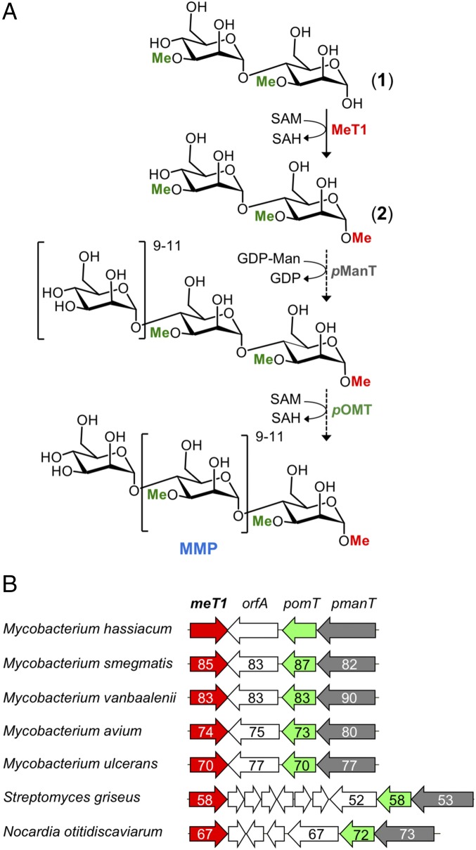
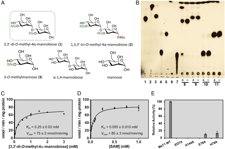
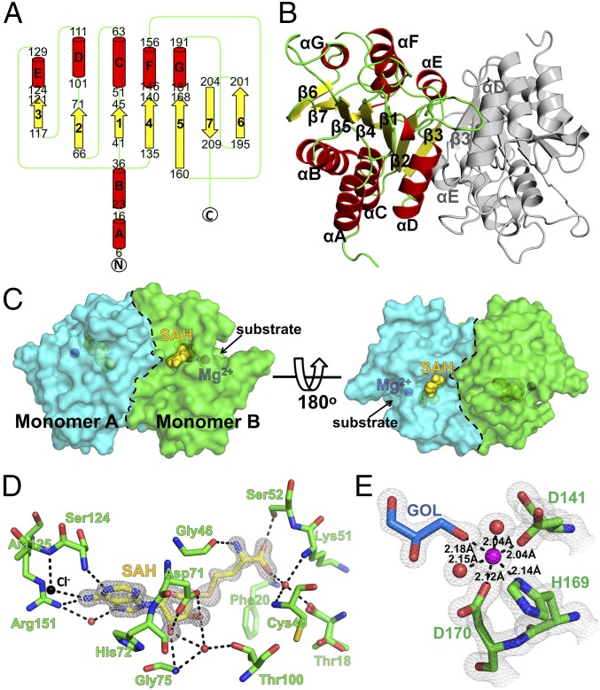
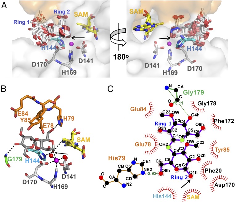
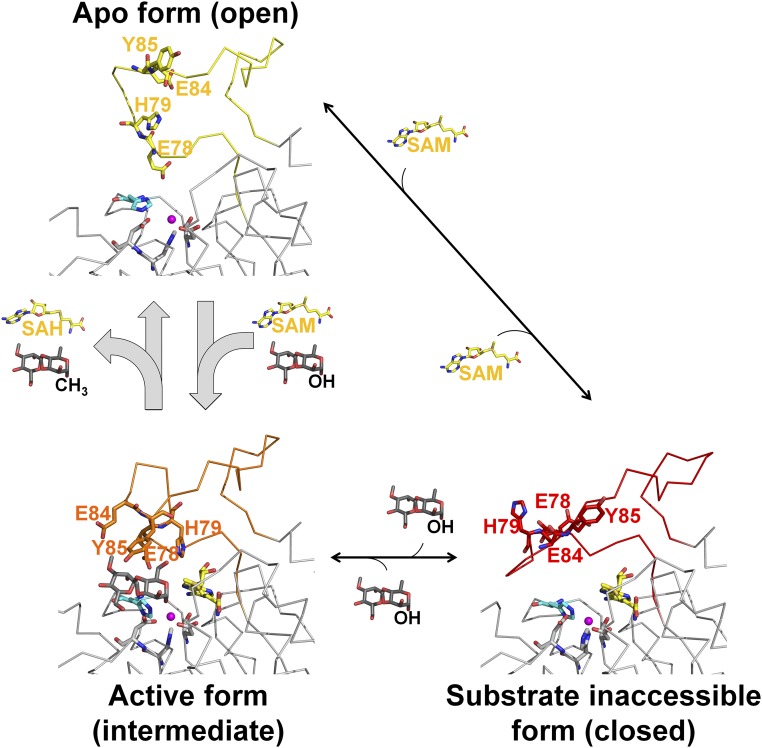
Mycobacteria are a wide group of organisms that includes strict pathogens, such as Mycobacterium tuberculosis, as well as environmental species known as nontuberculous mycobacteria (NTM), some of which-namely Mycobacterium avium-are important opportunistic pathogens. In addition to a distinctive cell envelope mediating critical interactions with the host immune system and largely responsible for their formidable resistance to antimicrobials, mycobacteria synthesize rare intracellular polymethylated polysaccharides implicated in the modulation of fatty acid metabolism, thus critical players in cell envelope assembly. These are the 6-O-methylglucose lipopolysaccharides (MGLP) ubiquitously detected across the Mycobacterium genus, and the 3-O-methylmannose polysaccharides (MMP) identified only in NTM. The polymethylated nature of these polysaccharides renders the intervening methyltransferases essential for their optimal function. Although the knowledge of MGLP biogenesis is greater than that of MMP biosynthesis, the methyltransferases of both pathways remain uncharacterized. Here, we report the identification and characterization of a unique S-adenosyl-l-methionine-dependent sugar 1-O-methyltransferase (MeT1) from Mycobacterium hassiacum that specifically blocks the 1-OH position of 3,3'-di-O-methyl-4α-mannobiose, a probable early precursor of MMP, which we chemically synthesized. The high-resolution 3D structure of MeT1 in complex with its exhausted cofactor, S-adenosyl-l-homocysteine, together with mutagenesis studies and molecular docking simulations, unveiled the enzyme's reaction mechanism. The functional and structural properties of this unique sugar methyltransferase further our knowledge of MMP biosynthesis and provide important tools to dissect the role of MMP in NTM physiology and resilience.

Keywords: 3D structure; Mycobacterium; S-adenosyl-l-methionine; polymethylated polysaccharides; sugar methyltransferase.


Conflict of interest statement

The authors declare no conflict of interest.

Figures

Fig. 1.
Fig. 1.
Proposed biosynthetic pathway of MMP and gene clusters encoding the intervening enzymes. (A) Proposed pathway for MMP biosynthesis in M. hassiacum with schematic representation of M. smegmatis MMP final product highlighting 3-O-methylation (green) and 1-O-methylation (red). Republished with permission of Royal Society of Chemistry, from ref. ; permission conveyed through Copyright Clearance Center, Inc. Dashed arrows represent inferred enzyme activities. (B) Organization of the proposed MMP biosynthetic genomic cluster in mycobacteria and related actinobacteria. meT1 (red), 1-O-methyltransferase; orfA (white), protein of unknown function; pomT (green), putative 3-O-methyltransferase; pmanT (gray), probable mannosyltransferase (19). The numbers indicate the level of amino acid identity (%) to the M. hassiacum orthologs.
Fig. 2.
Fig. 2.
Biochemical characterization of M. hassiacum MeT1. (A) Chemical structures of mannosides assayed as potential MeT1 substrates. (B) TLC analysis of MeT1 activity using SAM as methyl donor. Lanes 1–7, standards; lanes 8–11, MeT1 substrate assessment reactions, with the presence of enzyme denoted by (+) and the negative controls without enzyme by (−). Lane 1, synthetic 3,3-dimethylmannobiose (1); lane 2, synthetic 1,3,3-trimethylmannobiose (2); lane 3, synthetic 3-O-methylmannose (3); lane 4, α-1,4-mannobiose; lane 5, mannose; lane 6, SAM; lane 7, SAH; lane 8, reaction with MeT1 and compound 1; lane 9, reaction with MeT1 and compound 3; lane 10, reaction with MeT1 and α-1,4-mannobiose; lane 11, reaction with MeT1 and mannose. (C) Michaelis–Menten curve at 37 °C for 3,3-dimethylmannobiose (1) (in the presence of 1 mM SAM). (D) Michaelis–Menten curve at 37 °C for SAM [in the presence of 3.5 mM 3,3-dimethylmannobiose (1)]. (E) Effect of EDTA and of three key single amino acid substitutions on the catalytic activity of MeT1. Error bars represent SEM (C and D) or SD (E).
Fig. 3.
Fig. 3.
Three-dimensional structure and cofactor binding region of M. hassiacum MeT1. (A) Topology diagram of the MeT1 monomer. Alpha-helices (A–G) and β-strands (1–7) are represented as red cylinders and yellow arrows, respectively. The number of the first and last residue of each secondary structure element is indicated. (B) Cartoon representation of the MeT1 dimer, with the secondary structure elements of one of the monomers colored as in A and the other represented in gray. (C) Solid surface representation of the MeT1 dimer. Two pockets (one per monomer: monomer A, cyan; monomer B, green) in MeT1 allow substrate access to the active site. The blue and yellow spheres represent the magnesium ion and the SAH cofactor, respectively. A black dashed line indicates the dimer interface. The Right view results from a 180° rotation of the Left pose around x. (D) Close-up view of the extensive network of interactions established between the SAH moiety (stick model with: nitrogen atoms, blue; oxygen, red; and carbon, yellow) and MeT1 (only residues interacting with SAH are shown as sticks with carbon atoms green). The 2FoFc electron density map (3σ cut-off) for SAH is displayed as a gray mesh and black and red spheres represent chloride ions and water molecules, respectively. Hydrogen bonds are indicated by dashed black lines. (E) A magnesium ion is present at the active site of M. hassiacum MeT1. The triad formed by residues Asp141, His169, and Asp170, two water molecules and a glycerol molecule coordinate the active site Mg2+ of MeT1. The gray grid represents the 2FoFc electron density map (2σ cut-off). The protein residues and the glycerol molecule are represented as sticks with carbon atoms green (protein) or light blue (glycerol). The magnesium ion and water molecules are represented as magenta and red spheres, respectively. All coordination distances (black dashed lines) are indicated.
Fig. 4.
Fig. 4.
In silico substrate docking to MeT1. (A) Models of the quaternary complexes formed between MeT1, SAM, Mg2+, and 3,3-dimethylmannobiose (1). The enzyme is shown as a transparent white surface with the lid region colored orange (in the intermediate conformation, as depicted in SI Appendix, Fig. S14). The cofactor, the Mg2+-coordinating residues, and His144 are represented as yellow, gray, and cyan sticks, respectively, with oxygen atoms (red) and nitrogen (blue). All “productive” poses from the same positional cluster are displayed as white sticks, except for the lowest energy pose, gray. The magnesium ion is represented as a magenta sphere and the glycerol molecule as blue sticks. (Right) Results from a 180° rotation of the Left pose around y. (B) Close-up view of the interactions predicted for the lowest energy pose. In addition to the residues highlighted in A, Glu78, His79, Glu84, Tyr85, and Gly179, which interact with the disaccharide, are also represented as orange (if located in the lid region) or green sticks. Water molecules involved in Mg2+ coordination are represented as red spheres. (C) Two-dimensional ligand–protein interaction diagram for the lowest energy pose (drawn with LigPlot+). In all panels, a black arrow indicates the position of the methyl-accepting oxygen.
Fig. 5.
Fig. 5.
The lid domain of MeT1 adopts different conformations during its catalytic cycle. The open conformation of the lid, observed in the apo form of the enzyme (Upper) favors cofactor and substrate binding, which induces conformational changes leading to the intermediate lid conformation (Lower Left). In this intermediate state of the lid, residues Glu78 and His79 contact the 5-OH group of the methyl-accepting sugar ring and residues Glu84 and Tyr85 establish hydrophobic interactions with the 3′-O-methyl groups of the substrate, properly orienting it for processing. The 1-OH group of the constricted sugar ring coordinates the magnesium ion and is equidistant from the methyl group of SAM and the side chain of the catalytic base, His144. Once the methyl transfer reaction takes place, lid opening promotes product release and cofactor exchange, restarting the cycle. In the closed conformation of the lid (Lower Right), substrate access to the active site is impaired.

Similar articles

Cited by

References

    1. Falkinham JO., 3rd Environmental sources of nontuberculous mycobacteria. Clin Chest Med. 2015;36:35–41. - PubMed
    1. Koh WJ. Nontuberculous mycobacteria—Overview. Microbiol Spectr. 2017;5 - PMC - PubMed
    1. Gray GR, Ballou CE. Isolation and characterization of a polysaccharide containing 3-O-methyl-D-mannose from Mycobacterium phlei. J Biol Chem. 1971;246:6835–6842. - PubMed
    1. Ilton M, et al. Fatty acid synthetase activity in Mycobacterium phlei: Regulation by polysaccharides. Proc Natl Acad Sci USA. 1971;68:87–91. - PMC - PubMed
    1. Mendes V, Maranha A, Alarico S, Empadinhas N. Biosynthesis of mycobacterial methylglucose lipopolysaccharides. Nat Prod Rep. 2012;29:834–844. - PubMed

Publication types

MeSH terms

LinkOut - more resources