. 2019 Jan 18;363(6424):294-297.
doi: 10.1126/science.aau0583. Epub 2019 Jan 3.

H3K9me3-heterochromatin loss at protein-coding genes enables developmental lineage specification

Affiliations

H3K9me3-heterochromatin loss at protein-coding genes enables developmental lineage specification

Dario Nicetto et al. Science. .

Abstract

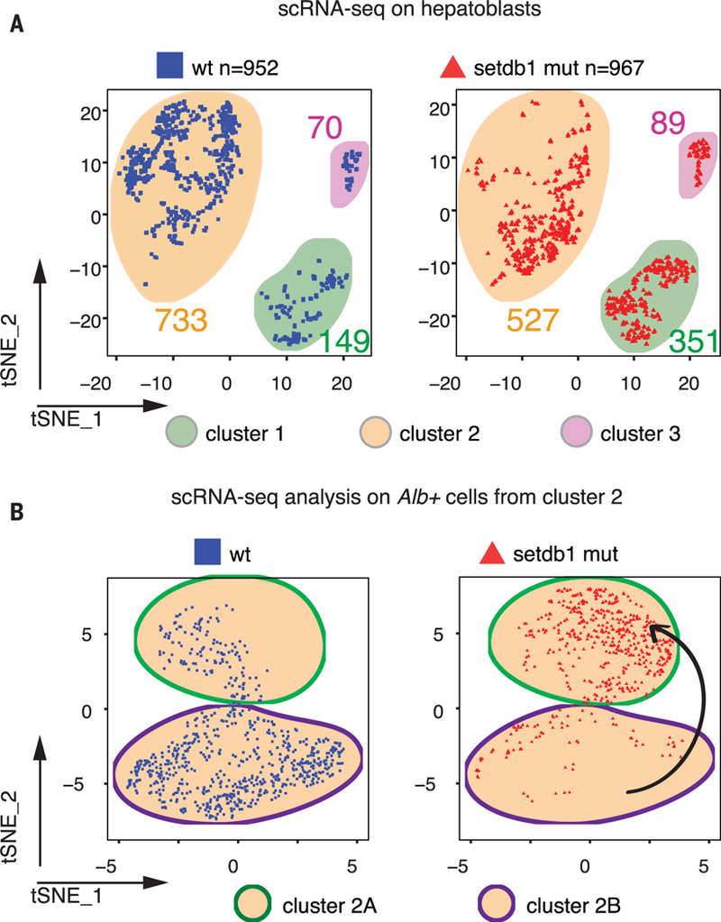
Gene silencing by chromatin compaction is integral to establishing and maintaining cell fates. Trimethylated histone 3 lysine 9 (H3K9me3)-marked heterochromatin is reduced in embryonic stem cells compared to differentiated cells. However, the establishment and dynamics of closed regions of chromatin at protein-coding genes, in embryologic development, remain elusive. We developed an antibody-independent method to isolate and map compacted heterochromatin from low-cell number samples. We discovered high levels of compacted heterochromatin, H3K9me3-decorated, at protein-coding genes in early, uncommitted cells at the germ-layer stage, undergoing profound rearrangements and reduction upon differentiation, concomitant with cell type-specific gene expression. Perturbation of the three H3K9me3-related methyltransferases revealed a pivotal role for H3K9me3 heterochromatin during lineage commitment at the onset of organogenesis and for lineage fidelity maintenance.


Figures

Fig. 1.
Fig. 1.. Chromatin compaction and H3K9me3 landscape upon germ-layer differentiation.
(A) Schematic of the cell types and embryonic developmental stages considered in this study. Purple, orange, and green asterisks next to the represented cell types indicate samples processed for H3K9me3 ChIP-seq, RNA-seq, and srHC-seq, respectively. (B) Alluvial plot showing dynamics in definitive endoderm srHC (dark gray) and open chromatin (light gray) patches upon differentiation into adult hepatocytes and insulin-producing cells; a to h indicate distinct categories or alluvia, characterized by a specific dynamic in srHC and open chromatin; the number of genes in each alluvium is indicated. (C)Number of genes marked by H3K9me3 in each indicated stage, along the hepatic and pancreatic lineages. Inner cell mass (ICM), e6.5, and e7.5 data from (25). (D)Number of genes gaining (purple) or losing (white) H3K9me3 upon stepwise transition in successive developmental stages. Each transition is indicated above the corresponding gene number bar.
Fig. 2.
Fig. 2.. Loss of srHC and H3K9me3 correlates with gene expression of hepatic-specific markers upon differentiation.
(A) Representative UCSC genome browser tracks of srHC-seq (gray), input-divided H3K9me3 (purple), and RNA-seq profiles (orange) upon definitive endoderm differentiation into adult hepatocytes. srHC and H3K9me3 patches are shown as gray and purple bars above each profile, respectively. Cytochrome P450 (Cyp) genes on chromosome 5 (A) are shown. The constitutive H3K9me3-undecorated and active Actin b (Actb) and the permanently H3K9me3-enriched and silenced zinc finger protein (Zfp) 936 (B) have been included as examples of genes whose expression inversely correlates to H3K9me3 presence. Magenta arrows indicate presence of srHC and H3K9me3 and absence of expression. Green arrows indicate absence of srHC, H3K9me3, and gene expression. (C) Z-score cluster representations for genes expressed in adult hepatocytes. (D and E) Heatmaps showing levels of srHC (D) and H3K9me3 (E) in the indicated stages. For both srHC and H3K9me3 heatmaps, definitive endodermal cell values have been ordered in a descendent manner.
Fig. 3.
Fig. 3.. Setdb1 mutant hepatoblasts up-regulate lineage-nonspecific genes.
(A) t-Distributed stochastic neighbor embedding (tSNE) plots of single-cell RNA-seq data showing wt (cells from n = 7 embryos; left, blue squares) and Setdb1 mutant (cells from n = 3 embryos; right, red triangles) cells in the three identified clusters. (B) tSNE plots of single cell RNA-seq data showing wt (top, blue squares) and Setdb1 mutant (bottom, red triangles) Albumin-positive cells from cluster 2. The black arrow indicates transition of Setdb1 mutant cells (red triangles) to a different cluster from that of the wt cells (blue squares).
Fig. 4.
Fig. 4.. TKO mutant cells lose hepatic identity and show developmental phenotypes associated with decreased H3K9me3 and srHC levels.
(A) tSNE plots of e11.5 single-cell RNA-seq data showing wt, Setdb1 mutant (same as in Fig. 3), and TKO (cells from n = 7 embryos; right, dark green circles) cells in the four identified clusters. (B) Representative morphological phenotype of 1-month-old control (ctrl) (n = 3) and FoxA3-cre; Setdb1 fl/fl; Suv39h1 fl/fl, Suv39h2 KO KO triple-knockout (TKO) mutants (n = 5). (C) Hematoxylin and eosin (H&E) staining and cytokeratin 7 immunohistochemistry in 1-month-old ctrl and TKO livers. Scale bar: 50 mm. (D) Percentage of genome covered by H3K9me3 and srHC domains in ctrl and TKO livers. (E) Representative electron microscopy images for ctrl and TKO 1-month-old hepatocytes. Scale bar: 600 nm. The number of cells recorded in the two groups is indicated at the bottom.

References

    1. Duboule D, Dev. Suppl, 135–142 (1994). - PubMed
    1. Raff RA, The Shape of Life: Genes, Development, and the Evolution of Animal Form (University of Chicago Press, IL, 1996).
    1. Irie N, Kuratani S, Development 141, 4649–4655 (2014). - PubMed
    1. Meshorer E, Misteli T, Nat. Rev. Mol. Cell Biol. 7, 540–546 (2006). - PubMed
    1. Wen B, Wu H, Shinkai Y, Irizarry RA, Feinberg AP, Nat. Genet. 41, 246–250 (2009). - PMC - PubMed

Publication types