Poxvirus infection in a Steller's sea eagle (Haliaeetus pelagicus)
- PMID: 30606906
- PMCID: PMC6395218
- DOI: 10.1292/jvms.18-0566
Poxvirus infection in a Steller's sea eagle (Haliaeetus pelagicus)
Abstract
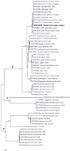
A severely emaciated adult Steller's sea eagle (Haliaeetus pelagicus) was found dead with electrocution-induced severe wing laceration, and with multiple cutaneous pock nodules at the periocular regions of both sides nearby the medial canthi and rhamphotheca. Histopathological examination of the nodules revealed hyperplasia of the epidermis with vacuolar degeneration and intracytoplasmic inclusion bodies (Bollinger bodies). The proventriculus was severely affected by nematodes and was ulcerated. Nucleotide sequencing of a PCR-amplified product of Avipoxvirus 4b core gene revealed 100% identity to the sequence of Avipoxvirus derived from other eagle species. This report describes the first detection of Avipoxvirus clade A from a Steller's sea eagle.
Keywords: 4b gene; Avipoxvirus; Japan; Steller’s sea eagle.
Figures


Similar articles
-
Avian poxvirus infection in a white-tailed sea eagle (Haliaeetus albicilla) in Japan.Avian Pathol. 2009 Dec;38(6):485-9. doi: 10.1080/03079450903349246. Avian Pathol. 2009. PMID: 19937537
-
Characterization of an Avipoxvirus From a Bald Eagle ( Haliaeetus leucocephalus ) Using Novel Consensus PCR Protocols for the rpo147 and DNA-Dependent DNA Polymerase Genes.J Avian Med Surg. 2016 Dec;30(4):378-385. doi: 10.1647/2015-120. J Avian Med Surg. 2016. PMID: 28107076
-
Avian pox infection in a free-living crested serpent eagle (Spilornis cheela) in southern Taiwan.Avian Dis. 2011 Mar;55(1):143-6. doi: 10.1637/9510-082610-Case.1. Avian Dis. 2011. PMID: 21500652
-
Multiple gene typing and phylogeny of avipoxvirus associated with cutaneous lesions in a stone curlew.Vet Res Commun. 2017 Jun;41(2):77-83. doi: 10.1007/s11259-016-9674-5. Epub 2017 Jan 4. Vet Res Commun. 2017. PMID: 28054222
-
Avipoxviruses: infection biology and their use as vaccine vectors.Virol J. 2011 Feb 3;8:49. doi: 10.1186/1743-422X-8-49. Virol J. 2011. PMID: 21291547 Free PMC article. Review.
References
-
- Abdo W., Magouz A., El-Khayat F., Kamal T.2017. Acute outbreak of co-infection of fowl pox and infectious laryngotracheitis viruses in chicken in Egypt. Pak. Vet. J. 37: 321–325.
-
- Cooper J. E.1969. Two cases of pox in recently imported peregrine falcons (Falco peregrinus). Vet. Rec. 85: 683–685. - PubMed

