Diagnostic Value of Contrast-Enhanced Digital Mammography versus Contrast-Enhanced Magnetic Resonance Imaging for the Preoperative Evaluation of Breast Cancer
- PMID: 30607168
- PMCID: PMC6310721
- DOI: 10.4048/jbc.2018.21.e62
Diagnostic Value of Contrast-Enhanced Digital Mammography versus Contrast-Enhanced Magnetic Resonance Imaging for the Preoperative Evaluation of Breast Cancer
Abstract
Purpose: This study aimed to compare the diagnostic performance of contrast-enhanced digital mammography (CEDM) and contrast-enhanced magnetic resonance imaging (CEMRI) in preoperative evaluations, and to evaluate the effect of each modality on the surgical management of women with breast cancer.
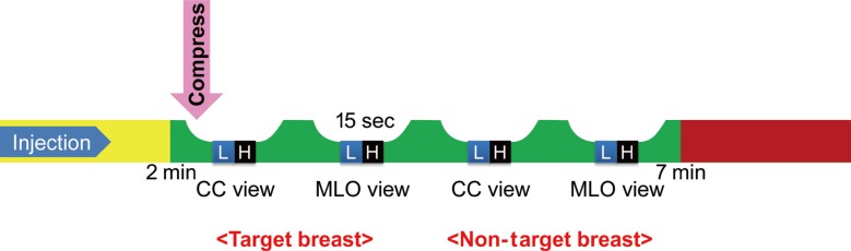
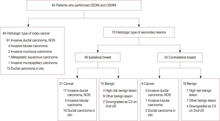
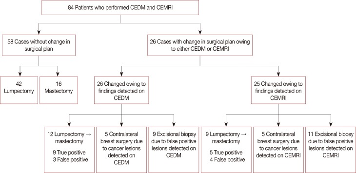
Methods: This single-center, prospective study was approved by the Institutional Review Board, and informed consent was obtained from all patients. From November 2016 to October 2017, 84 patients who were diagnosed with invasive carcinoma (69/84) and ductal carcinoma in situ (15/84), and underwent both CEDM and CEMRI, were enrolled. Imaging findings and surgical management were correlated with pathological results and compared. The diagnostic performance of both modalities in the detection of index and secondary cancers (multifocality and multicentricity), and occult cancer in the contralateral breast, was compared. The authors also evaluated whether CEDM or CEMRI resulted in changes in the surgical management of the affected breast due to imaging-detected findings.
Results: Eighty-four women were included in the analysis. Compared with CEMRI, CEDM demonstrated a similar sensitivity (92.9% [78/84] vs. 95.2% [80/84]) in detecting index cancer (p=0.563). For the detection of secondary cancers in the ipsilateral breast and occult cancer in the contralateral breast, no significant differences were found between CEDM and CEMRI (p=0.999 and p=0.999, respectively). Regarding changes in surgical management, CEDM resulted in similar changes compared with CEMRI (30.9% [26/84] vs. 29.7% [25/84], p=0.610). Regarding changes in surgical management due to false-positive findings, no significant differences were found between CEDM and CEMRI (34.6% [9/26] vs. 44.0% [11/25], p=0.782).
Conclusion: CEDM demonstrated a diagnostic performance comparable with CEMRI in depicting index cancers, secondary cancers, and occult cancer in the contralateral breast. CEDM demonstrated similar changes in surgical management compared with CEMRI.
Keywords: Breast neoplasms; Contrast media; Magnetic resonance imaging; Mammography.
Conflict of interest statement
CONFLICT OF INTEREST: The authors declare that they have no competing interests.
Figures
References
-
- Miller BT, Abbott AM, Tuttle TM. The influence of preoperative MRI on breast cancer treatment. Ann Surg Oncol. 2012;19:536–540. - PubMed
-
- Houssami N, Hayes DF. Review of preoperative magnetic resonance imaging (MRI) in breast cancer: should MRI be performed on all women with newly diagnosed, early stage breast cancer? CA Cancer J Clin. 2009;59:290–302. - PubMed
-
- Houssami N, Ciatto S, Macaskill P, Lord SJ, Warren RM, Dixon JM, et al. Accuracy and surgical impact of magnetic resonance imaging in breast cancer staging: systematic review and meta-analysis in detection of multifocal and multicentric cancer. J Clin Oncol. 2008;26:3248–3258. - PubMed
-
- Lewin JM, Isaacs PK, Vance V, Larke FJ. Dual-energy contrast-enhanced digital subtraction mammography: feasibility. Radiology. 2003;229:261–268. - PubMed
LinkOut - more resources
Full Text Sources