. 2018 Mar 22;6(1):4.
doi: 10.1007/s40203-018-0041-9. eCollection 2018.

Sulfonamide derivatives as Mycobacterium tuberculosis inhibitors: in silico approach

Affiliations

Sulfonamide derivatives as Mycobacterium tuberculosis inhibitors: in silico approach

Sayantan Pradhan et al. In Silico Pharmacol. .

Abstract

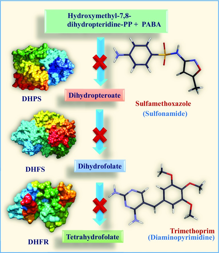
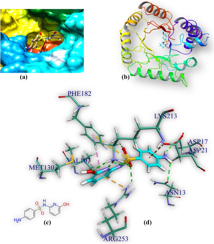
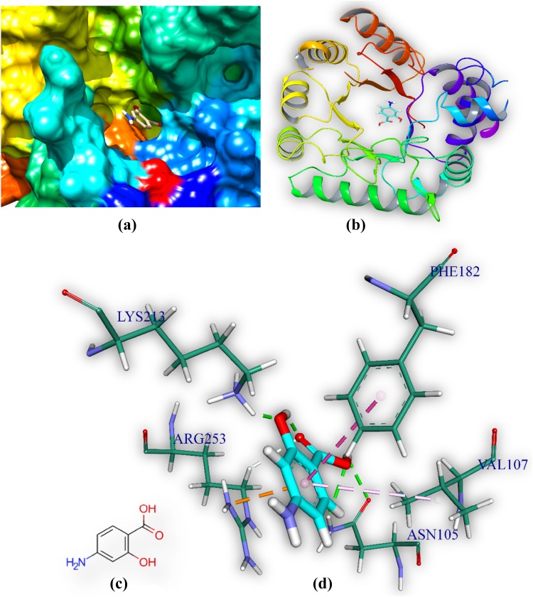
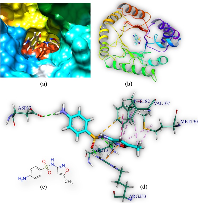
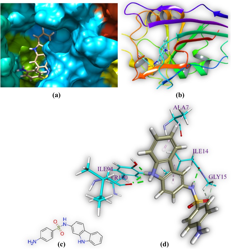
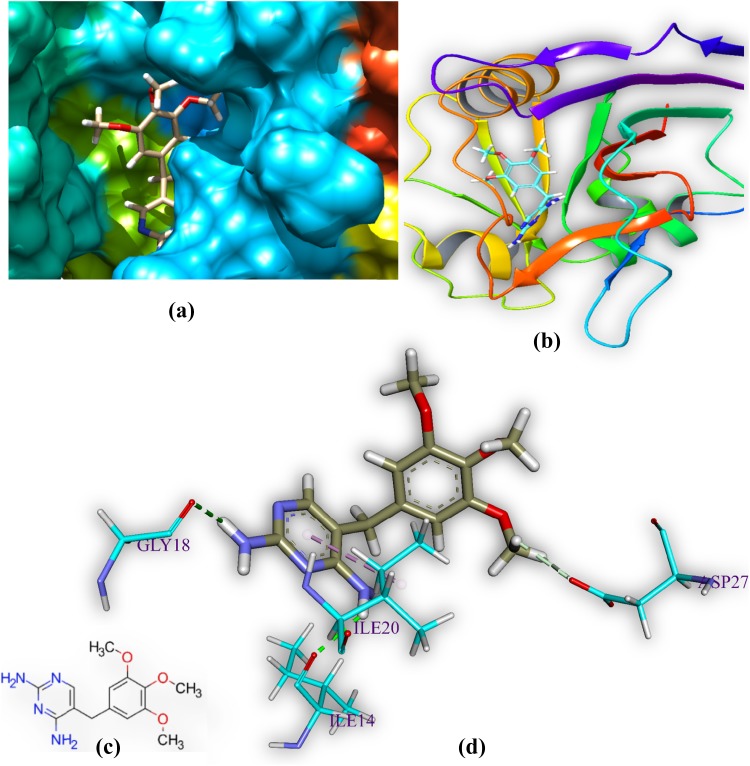
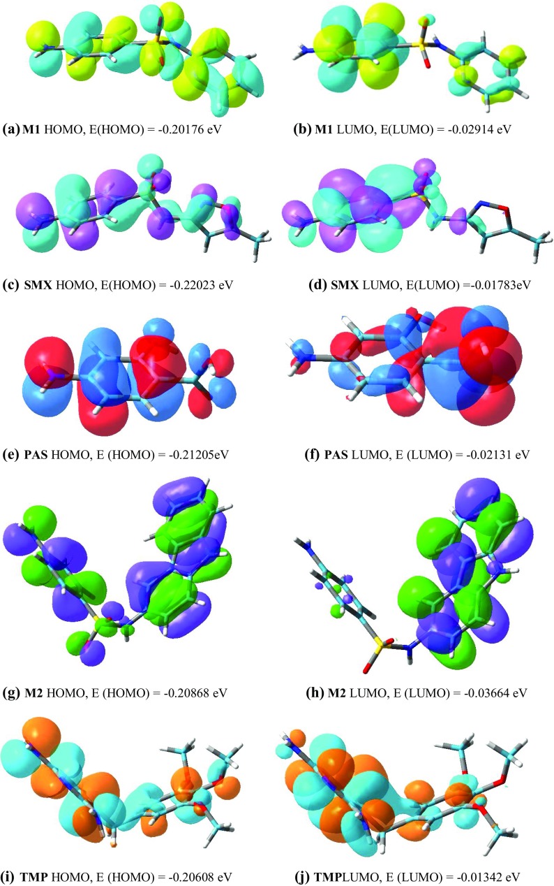
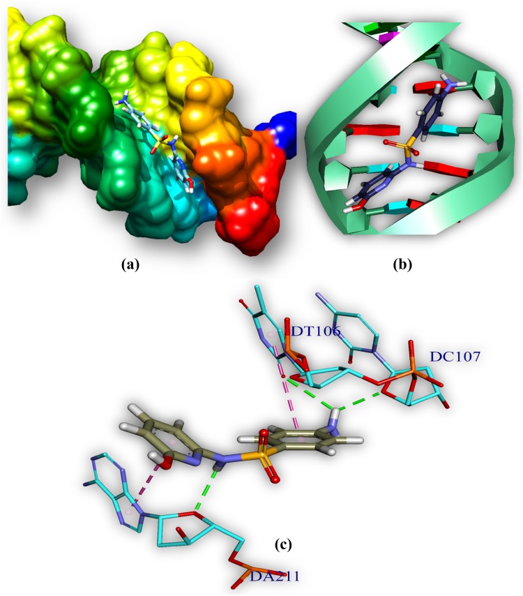
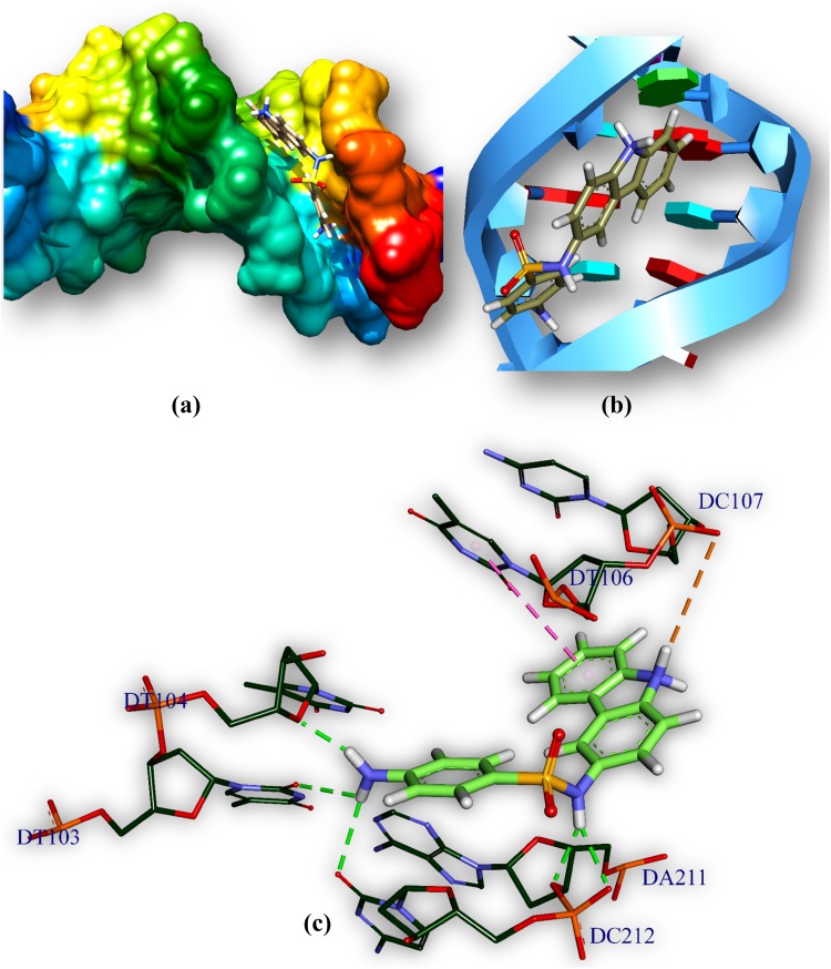
Both DHPS (dihydropteroate synthase) and DHFR (dihydrofolate reductase) play important physiological roles in the survivability of Mycobacterium tuberculosis (MTB). Sulfonamides are the potent drugs to monitor growth and proliferation of MTBs by inhibiting the activity of DHPS and DHFR which could explain the mechanism of action of these molecules. In this work, 102 heterocyclic sulfonamides (HSF) have been screened by discovery studio molecular docking programme to search the best suitable molecule for the treatment of MTBs. Lipinski's rule of five protocols is followed to screen drug likeness of these molecules and ADMET (absorption, distribution, metabolism, excretion and toxicity) filtration has been used to value their toxicity. Only fourteen molecules are found to obey the Lipinski's rule and able to cross the ADMET filter. A small difference between HOMO and LUMO energy signifies the electronic excitation energy which is essential to calculate molecular reactivity and stability of the best docked compound and easy activation of drug in the protein environment. Both 4-amino-N-(6-hydroxypyridin-2-yl)benzenesulfonamide (M1) and 4-amino-N-(9H-carbazol-2-yl)benzenesulfonamide (M2) show the best theoretical efficiency with DHPS and DHFR, respectively. These compounds are also found to bind to the adenine-thymine region of tuberculosis DNA.

Keywords: ADMET; DHPS and DHFR inhibitors; Heterocyclic sulfonamide compounds; Molecular docking; Structure based drug design.


Conflict of interest statement

Compliance with ethical standardsThe authors confirm that this article content has no conflicts of interest.

Figures

Fig. 1
Fig. 1
Mode of action of sulfonamides on Tetrahydrofolate synthesis pathway
Fig. 2
Fig. 2
Protein (DHFR) multiple sequence alignment (Jalview) generated by Clustal omega (partial view). The amino acid sequence of the human DHFR protein has been compared with various M. tuberculosis DHFR. Site specific similarities between amino acids are shown by the colored letters while the degree of conservation, quality and consensus of those amino acids among all organisms are shown in yellow and black
Fig. 3
Fig. 3
Comprehensive perception of DHPS and M1 after docking, a secondary structure of MTB DHPS represented by hydrophobic surface and M1 represented is by stick model and coloured according to elements, b DHPS is represented by ribbon and M1 is represented by stick, c 2D structure of M, and d interactions of M1 with DHPS amino acids. Bonds are in dots. M1 surrounding amino acids are in three letters code, represented in blue. For brevity explicit H-atoms are deleted in 2D structure
Fig. 4
Fig. 4
Comprehensive perception of DHPS and PAS after docking. Figure legends are same as Fig. 3
Fig. 5
Fig. 5
Comprehensive perception of DHPS and sulfamethoxazole after docking. Figure legends are same as Fig. 3
Fig. 6
Fig. 6
Comprehensive perception of DHFR and M2 after docking. Figure legends are same as Fig. 3
Fig. 7
Fig. 7
Comprehensive perception of DHFR and trimethoprim after docking. Figure legends are same as Fig. 3
Fig. 8
Fig. 8
HOMO and LUMO of M1, SMX, PAS, M2 and TPM
Fig. 9
Fig. 9
Comprehensive perception of MTB DNA and M1 after docking. M1 bound to MTB DNA in minor groove, a double helical structure of DNA represented by hydrophobic surface and M1 is represented by stick model and are coloured according to elements, b M1 is represented by stick and double helical DNA structures is represented by ladder and rings, c interactions of ligand with DNA base pairs (A, T, G, C), the interaction type. Bonds are in dots, ligand surrounding base pairs are in two letter code represented in blue
Fig. 10
Fig. 10
Comprehensive perception of MTB DNA and M2 after docking. Figure legends are same as Fig. 9

Similar articles

Cited by

References

    1. Ajay, Bemis GW, Murcko MA. Designing libraries with CNS activity. J Med Chem. 1999;42(24):4942–4951. doi: 10.1021/jm990017w. - DOI - PubMed
    1. Ames BN, Gurney EG, Miller JA, Bartsch H. Carcinogens as frameshift mutagens: metabolites and derivatives of 2-acetylaminofluorene and other aromatic amine carcinogens. Proc Natl Acad Sci USA. 1972;69(11):3128–3132. doi: 10.1073/pnas.69.11.3128. - DOI - PMC - PubMed
    1. Becke AD. Density-functional thermochemistry. I. The role of exact exchange. J Chem Phys. 1993;98(7):5648–5652. doi: 10.1063/1.464913. - DOI
    1. Cheah H-L, Lim V, Sandai D. Inhibitors of the glyoxylate cycle enzyme ICL1 in Candida albicans for potential use as antifungal agents. PLoS One. 2014;9(4):e95951. doi: 10.1371/journal.pone.0095951. - DOI - PMC - PubMed
    1. Cheng F, Li W, Zhou Y, Shen J, Wu Z, Liu G, Lee PW, Tang Y. admetSAR: a comprehensive source and free tool for assessment of chemical ADMET properties. J Chem Inf Model. 2012;52(11):3099–3105. doi: 10.1021/ci300367a. - DOI - PubMed

LinkOut - more resources