Case Reports
. 2018 Dec;48(4):289-293.
doi: 10.5624/isd.2018.48.4.289. Epub 2018 Dec 20.

Giant complex odontoma in the posterior mandible: A case report and literature review

Affiliations
Case Reports

Giant complex odontoma in the posterior mandible: A case report and literature review

Jong Chan Park et al. Imaging Sci Dent. 2018 Dec.

Abstract

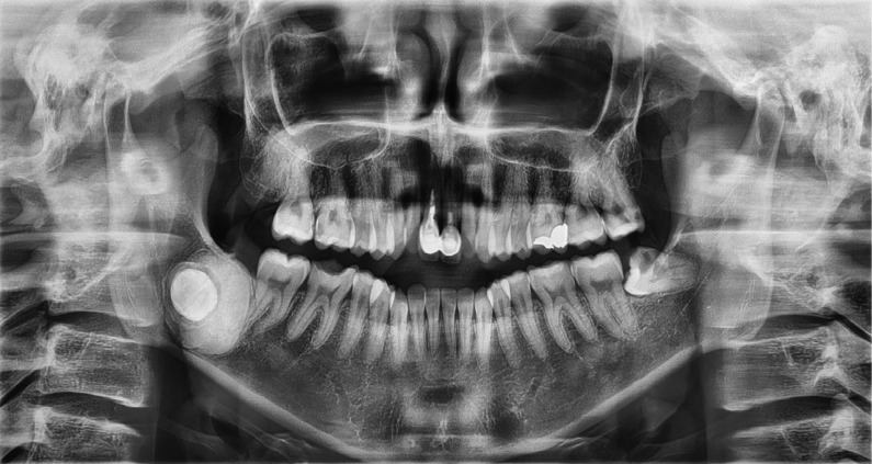
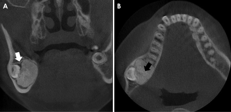
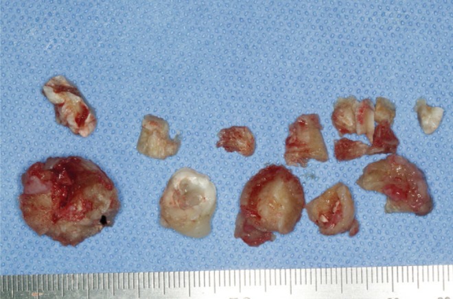
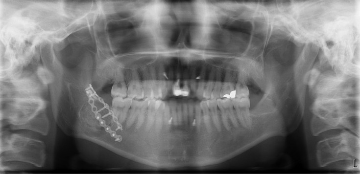
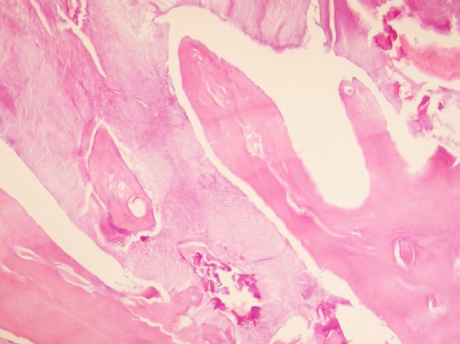
Odontomas are considered a type of odontogenic hamartoma, and are generally reported not to exceed 3 cm in diameter. Some authors have referred to odontomas with a diameter exceeding 3 cm as giant odontomas. As hamartomas, giant odontomas generally show no signs or symptoms, but if they perforate the mucosa to become exposed in the oral cavity, oral and maxillofacial infections can result. Surgical removal and a histopathological examination may also be required to differentiate them from osteomas, cemento-osseous dysplasia, or mixed odontogenic tumors. This report presents the case of a 28-year-old woman with a giant odontoma in the right mandibular third molar area. Based on a review of the literature published since 2010, only 11 cases of "giant" or "large" odontomas have been reported, most of which were of the complex odontoma type. It was confirmed that they tend to occur in the right posterior mandible.

Keywords: Hamartoma; Odontogenic Tumors; Odontoma; Radiography.


Figures

Fig. 1
Fig. 1. Panoramic radiograph taken on the first visit. The radiopaque lesion can be observed around the impacted right mandibular third molar.
Fig. 2
Fig. 2. Coronal and axial computed tomography views taken on the first visit. A. The radiopaque lesion is surrounded by a narrow radiolucent rim and dental follicle of the impacted right mandibular third molar. The radiolucent lesion of the impacted third molar is continuous with the radiolucent rim of the giant odontoma (white arrow). B. A 20×30×25 mm radiopaque lesion is observed on the right side of mandible. Expansion and thinning of the buccal cortical bone are observed on the right side of the mandible. The heterogenous radiopacity of the mass is also visible on the radiograph (black arrow).
Fig. 3
Fig. 3. Clinical photograph showing fragments of a complex odontoma with a left mandibular impacted third molar.
Fig. 4
Fig. 4. A postoperative panoramic image shows that the mass had been removed, the left maxillary and mandibular third molars and the right mandibular third molar had been extracted, and the left iliac block bone had been fixed with mini-plates. Maxillomandibular fixation was performed using a skeletal anchorage system screw and rubber.
Fig. 5
Fig. 5. Decalcified section of an odontoma shows a disorganized mass of randomly-arranged dentin intermixed with enamel matrix (hematoxylin and eosin staining, ×100).

Similar articles

Cited by

References

    1. Bagewadi SB, Kukreja R, Suma GN, Yadav B, Sharma H. Unusually large erupted complex odontoma: a rare case report. Imaging Sci Dent. 2015;45:49–54. - PMC - PubMed
    1. Hidalgo-Sánchez O, Leco-Berrocal MI, Martínez-González JM. Metaanalysis of the epidemiology and clinical manifestations of odontomas. Med Oral Patol Oral Cir Bucal. 2008;13:E730–E734. - PubMed
    1. An SY, An CH, Choi KS. Odontoma: a retrospective study of 73 cases. Imaging Sci Dent. 2012;42:77–81. - PMC - PubMed
    1. Spini PH, Spini TH, Servato JP, Faria PR, Cardoso SV, Loyola AM. Giant complex odontoma of the anterior mandible: report of case with long follow up. Braz Dent J. 2012;23:597–600. - PubMed
    1. Perumal CJ, Mohamed A, Singh A, Noffke CE. Sequestrating giant complex odontoma: a case report and review of the literature. J Maxillofac Oral Surg. 2013;12:480–484. - PMC - PubMed

Publication types