

Schwann cells shape the neuro-immune environs and control cancer progression
- PMID: 30607548
- PMCID: PMC11028256
- DOI: 10.1007/s00262-018-02296-3
Schwann cells shape the neuro-immune environs and control cancer progression
Abstract
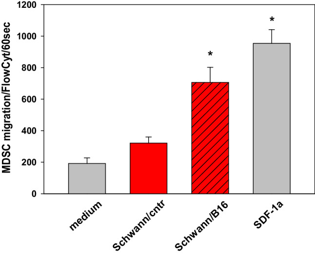
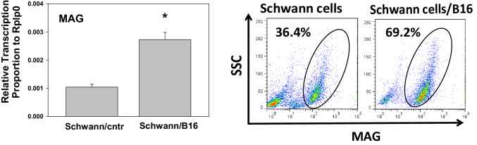
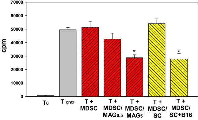
At present, significant experimental and clinical data confirm the active involvement of the peripheral nervous system (PNS) in different phases of cancer development and progression. Most of the research effort focuses on the impact of distinct neuronal types, e.g., adrenergic, cholinergic, dopaminergic, etc. in carcinogenesis, generally ignoring neuroglia. The very fact that these cells far outnumber the other cellular types may also play an important role worthy of study in this context. The most prevalent neuroglia within the PNS consists of Schwann cells (SCs). These cells play a substantial role in maintaining homeostasis within the nervous system. They possess distinct immunomodulatory, inflammatory and regenerative capacities-also, one should consider their broad distribution throughout the body; this makes them a perfect target for malignant cells during the initial stages of cancer development and the very formation of the tumor microenvironment itself. We show that SCs in the tumor milieu attract different subsets of immune regulators and augment their ability to suppress effector T cells. SCs may also up-regulate invasiveness of tumor cells and support metastatic disease. We outline the interactive potential of SCs juxtaposed with cancerous cells, referring to data from various external sources alongside data of our own.
Keywords: Cancer; MDSC; Neuroglia; PIVAC 18; Schwann cells; Tumor microenvironment.
Conflict of interest statement
The author reports no conflicts of interest in this work.
Figures
Similar articles
-
The Emerging Role of Schwann Cells in the Tumor Immune Microenvironment and Its Potential Clinical Application.Int J Mol Sci. 2024 Dec 23;25(24):13722. doi: 10.3390/ijms252413722. Int J Mol Sci. 2024. PMID: 39769484 Free PMC article. Review.
-
Tumor-neuroglia interaction promotes pancreatic cancer metastasis.Theranostics. 2020 Apr 6;10(11):5029-5047. doi: 10.7150/thno.42440. eCollection 2020. Theranostics. 2020. PMID: 32308766 Free PMC article.
-
Schwann cells: a new player in the tumor microenvironment.Cancer Immunol Immunother. 2017 Aug;66(8):959-968. doi: 10.1007/s00262-016-1929-z. Epub 2016 Nov 24. Cancer Immunol Immunother. 2017. PMID: 27885383 Free PMC article. Review.
-
Schwann Cells Augment Cell Spreading and Metastasis of Lung Cancer.Cancer Res. 2018 Oct 15;78(20):5927-5939. doi: 10.1158/0008-5472.CAN-18-1702. Epub 2018 Aug 22. Cancer Res. 2018. PMID: 30135194
-
Schwann Cells in Peripheral Cancers: Bystanders or Promoters?Adv Biol (Weinh). 2022 Sep;6(9):e2200033. doi: 10.1002/adbi.202200033. Epub 2022 Jun 3. Adv Biol (Weinh). 2022. PMID: 35656739 Review.
Cited by
-
Schwann cell plasticity regulates neuroblastic tumor cell differentiation via epidermal growth factor-like protein 8.Nat Commun. 2021 Mar 12;12(1):1624. doi: 10.1038/s41467-021-21859-0. Nat Commun. 2021. PMID: 33712610 Free PMC article.
-
The Emerging Role of Schwann Cells in the Tumor Immune Microenvironment and Its Potential Clinical Application.Int J Mol Sci. 2024 Dec 23;25(24):13722. doi: 10.3390/ijms252413722. Int J Mol Sci. 2024. PMID: 39769484 Free PMC article. Review.
-
Schwann cells in regeneration and cancer: an epithelial-mesenchymal transition perspective.Open Biol. 2025 Mar;15(3):240337. doi: 10.1098/rsob.240337. Epub 2025 Mar 5. Open Biol. 2025. PMID: 40037534 Free PMC article. Review.
-
Neuro-immune crosstalk in cancer: mechanisms and therapeutic implications.Signal Transduct Target Ther. 2025 Jun 2;10(1):176. doi: 10.1038/s41392-025-02241-8. Signal Transduct Target Ther. 2025. PMID: 40456735 Free PMC article. Review.
-
Novel Glial Cell Functions: Extensive Potency, Stem Cell-Like Properties, and Participation in Regeneration and Transdifferentiation.Front Cell Dev Biol. 2020 Aug 18;8:809. doi: 10.3389/fcell.2020.00809. eCollection 2020. Front Cell Dev Biol. 2020. PMID: 33015034 Free PMC article. Review.
References
Publication types
MeSH terms
Grants and funding
LinkOut - more resources
Full Text Sources
