

Potential usefulness of 68Ga-citrate PET/CT in detecting infected lower limb prostheses
- PMID: 30607646
- PMCID: PMC6318156
- DOI: 10.1186/s13550-018-0468-3
Potential usefulness of 68Ga-citrate PET/CT in detecting infected lower limb prostheses
Abstract
Background: Prosthetic joint infections may lead to failures of total joint arthroplasty. Radionuclide imaging can play a diagnostic role in identifying such infections, which require two-stage exchange arthroplasty (instead of simple revision surgery performed in non-infected cases). Although 18F-FDG PET/CT has emerged as a novel diagnostic tool in this setting, the clinical usefulness of 68Ga-citrate PET/CT has not been previously investigated. This single-center prospective study was designed to address this issue.
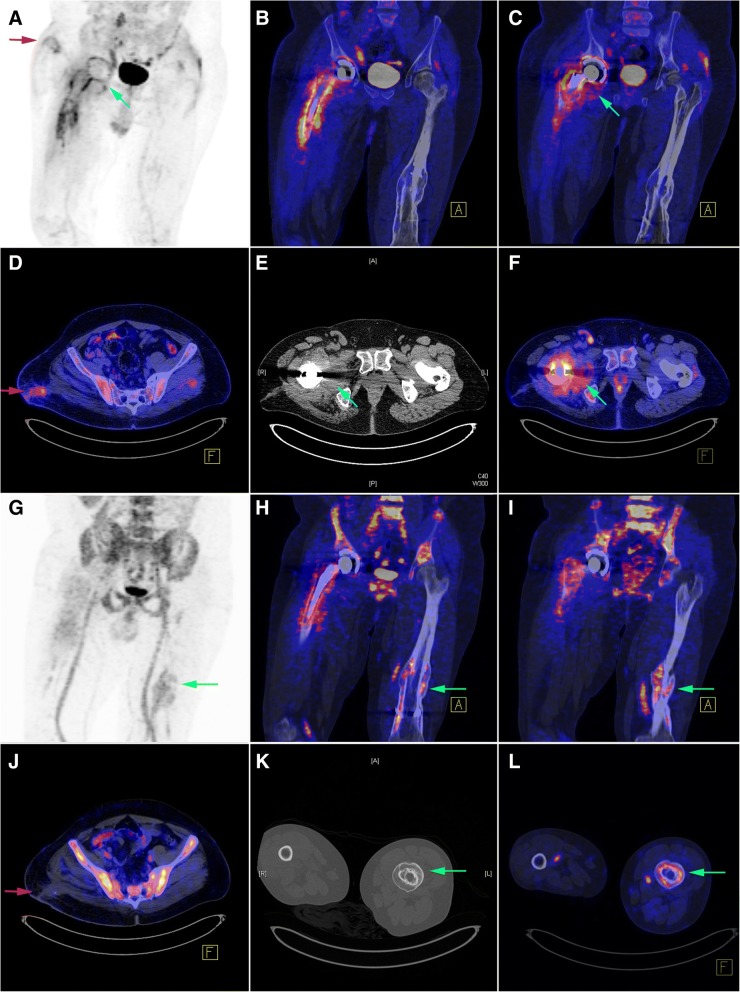
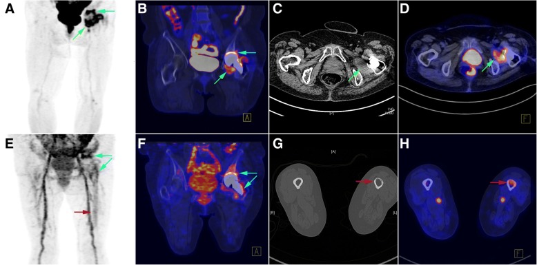
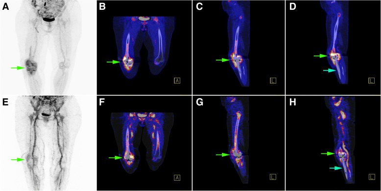
Methods: Between January 2016 and October 2017, we examined 34 patients with clinically proven or suspected prosthetic hip/knee joint infections scheduled to undergo surgery. All patients underwent 68Ga-citrate PET/CT scans and sequential 18F-FDG PET/CT imaging for comparative purposes. Intraoperative findings and the results of microbiological analyses of surgical specimens served as gold standard. The diagnostic results were examined according to (1) image interpretation based on radiotracer uptake patterns and (2) quantitative analysis using volumes of interest (VOIs) to calculate standard uptake values (SUVs) and metabolic volumes (MVs).
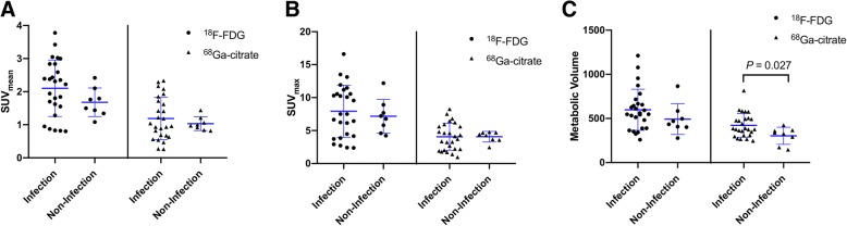
Results: A total of 26 (76%) patients were diagnosed as having infections. Based on radiotracer uptake pattern criteria, the sensitivity, specificity, and accuracy of 68Ga-citrate PET/CT and 18F-FDG PET/CT were 92%, 88%, and 91% and 100%, 38%, and 85%, respectively. MV was significantly higher in the infected group when 68Ga-citrate PET/CT was used (422.45 vs. 303.65 cm3, p = 0.027), whereas no significant differences were observed on 18F-FDG PET/CT. According to receiver operating characteristic (ROC) curve analysis, a cut-off value of 370.86 for MV resulted in a sensitivity of 61.5% and a specificity of 87.5% (area under curve: 0.75, 95% confidence interval: 0.57-0.88, p = 0.035).
Conclusions: Subject to future confirmation, our data provide preliminary evidence that 68Ga-citrate PET/CT may have a complimentary role to 18F-FDG PET/CT in detecting prosthetic joint infections, being characterized by a higher specificity and the possibility to discriminate between an infectious condition and sterile inflammation.
Trial registration: This prospective study was registered at clinicaltrials.gov (registration number: NCT02855190 ).
Keywords: 18F-FDG; 68Ga-citrate; PET/CT; Prosthetic joint infections.
Conflict of interest statement
Ethics approval and consent to participate
This prospective study was performed in accordance with the 1964 Declaration of Helsinki and was approved by the Institutional Review Board of the Chang Gung Memorial Hospital (CGMH) at Linkou (approval number: 103-7266A).
The study protocol was approved by the Institutional Review Board of the Chang Gung Memorial Hospital (CGMH) at Linkou.
Consent for publication
Written informed consent was obtained for all participants.
Competing interests
The authors declare that they have no competing interests.
Publisher’s Note
Springer Nature remains neutral with regard to jurisdictional claims in published maps and institutional affiliations.
Figures

Similar articles
-
More advantages in detecting bone and soft tissue metastases from prostate cancer using 18F-PSMA PET/CT.Hell J Nucl Med. 2019 Jan-Apr;22(1):6-9. doi: 10.1967/s002449910952. Epub 2019 Mar 7. Hell J Nucl Med. 2019. PMID: 30843003
-
PET/CT to detect adverse reactions to metal debris in patients with metal-on-metal hip arthroplasty: an exploratory prospective study.Clin Physiol Funct Imaging. 2018 Sep;38(5):847-855. doi: 10.1111/cpf.12493. Epub 2017 Dec 27. Clin Physiol Funct Imaging. 2018. PMID: 29280283
-
(68)Ga-radiopharmaceuticals for PET imaging of infection and inflammation.Recent Results Cancer Res. 2013;194:189-219. doi: 10.1007/978-3-642-27994-2_11. Recent Results Cancer Res. 2013. PMID: 22918761 Review.
-
The diagnostic accuracy of 18F-FDG PET/CT in diagnosing fracture-related infections.Eur J Nucl Med Mol Imaging. 2019 Apr;46(4):999-1008. doi: 10.1007/s00259-018-4218-6. Epub 2018 Dec 7. Eur J Nucl Med Mol Imaging. 2019. PMID: 30523391 Free PMC article.
-
Diagnostic Accuracy of 18F-FDG PET/CT in Infective Endocarditis and Implantable Cardiac Electronic Device Infection: A Cross-Sectional Study.J Nucl Med. 2016 Nov;57(11):1726-1732. doi: 10.2967/jnumed.116.173690. Epub 2016 Jun 3. J Nucl Med. 2016. PMID: 27261514 Review.
Cited by
-
Diagnostic accuracy of positron emission tomography/computerized tomography for periprosthetic joint infection of hip: systematic review and meta-analysis.J Orthop Surg Res. 2023 Aug 30;18(1):640. doi: 10.1186/s13018-023-04061-4. J Orthop Surg Res. 2023. PMID: 37644493 Free PMC article.
-
Fully Automated Macro- and Microfluidic Production of [68Ga]Ga-Citrate on mAIO® and iMiDEVTM Modules.Molecules. 2022 Feb 1;27(3):994. doi: 10.3390/molecules27030994. Molecules. 2022. PMID: 35164258 Free PMC article.
-
Medical imaging diagnosis of orthopedic prosthesis-associated infections: a narrative review.Quant Imaging Med Surg. 2025 Jan 2;15(1):947-961. doi: 10.21037/qims-24-403. Epub 2024 Nov 15. Quant Imaging Med Surg. 2025. PMID: 39839020 Free PMC article. Review.
-
Radionuclide Imaging of Fungal Infections and Correlation with the Host Defense Response.J Fungi (Basel). 2021 May 22;7(6):407. doi: 10.3390/jof7060407. J Fungi (Basel). 2021. PMID: 34067410 Free PMC article. Review.
-
68Ga-Citrate PET of Healthy Men: Whole-Body Biodistribution Kinetics and Radiation Dose Estimates.J Nucl Med. 2022 Oct;63(10):1598-1603. doi: 10.2967/jnumed.122.263884. Epub 2022 Mar 10. J Nucl Med. 2022. PMID: 35273093 Free PMC article.
References
Associated data
Grants and funding
LinkOut - more resources
Full Text Sources
Other Literature Sources
Medical
