. 2019 Jan 29;7(2):571-580.
doi: 10.1039/c8bm01253a.

Adipose-derived stem cell-secreted factors promote early stage follicle development in a biomimetic matrix

Affiliations

Adipose-derived stem cell-secreted factors promote early stage follicle development in a biomimetic matrix

Claire E Tomaszewski et al. Biomater Sci. .

Abstract

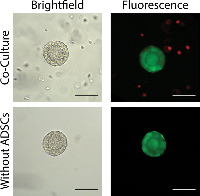
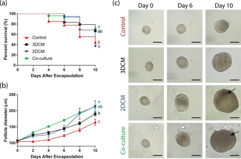
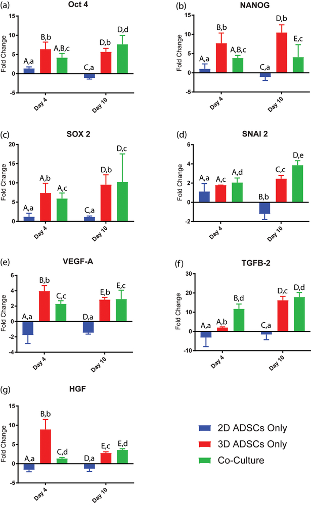
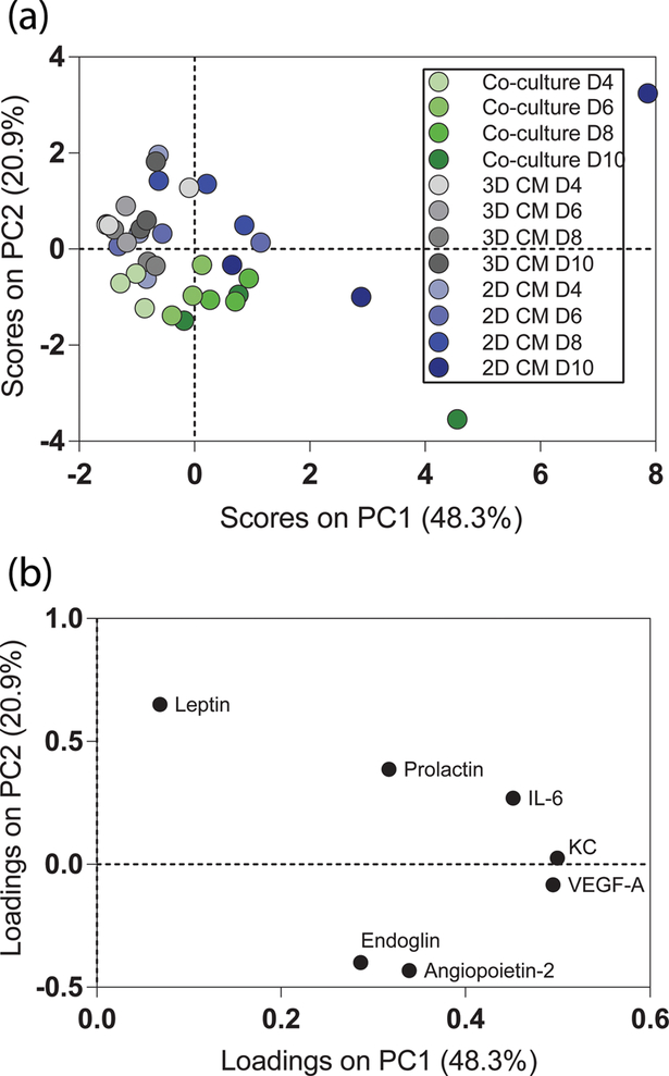
Development of primary follicles in vitro benefits from a three-dimensional matrix that is enriched with paracrine factors secreted from feeder cells and mimics the in vivo environment. In this study, we investigated the role of paracrine signaling from adipose-derived stem cells (ADSCs) in supporting primary follicle development in a biomimetic poly(ethylene glycol) (PEG)-based matrix. Follicles co-cultured with ADSCs and follicles cultured in conditioned medium from ADSCs encapsulated in gels (3D CM) exhibited significantly (p < 0.01 and p = 0.09, respectively) improved survival compared to follicles cultured in conditioned medium collected from ADSCs cultured in flasks (2D CM) and follicles cultured without paracrine support. The gene expression of ADSCs suggested that the stem cells maintained their multipotency in the 3D PEG environment over the culture period, regardless of the presence of the follicles, while under 2D conditions the multipotency markers were downregulated. The differences in cytokine signatures of follicles exposed to 3D and 2D ADSC paracrine factors suggest that early cytokine interactions are key for follicle survival. Taken together, the biomimetic PEG scaffold provides a three-dimensional, in vivo-like environment to induce ADSCs to secrete factors which promote early stage ovarian follicle development and survival.


Conflict of interest statement

Conflicts of Interest

The authors have no conflicts of interest to declare.

Figures

Fig 1.
Fig 1.. Fluorescent labeling of follicles co-cultured with and without adipose-derived stem cells.
GFP-labeled follicles (green) encapsulated in PEG hydrogels with and without (red) fluorescently labeled ADSCs on day 0 of culture (scale bars are 100μm). Without integrin-binding sequences, ADSCs remain isolated from the follicle when co-cultured in PEG.
Fig 2.
Fig 2.. Growth and survival of follicles co-cultured with ADSCs and follicles cultured in ADSC-conditioned medium.
(a) Survival and (b) growth of follicles co-cultured with ADSCs (green), cultured in conditioned medium from ADSCs cultured in a 3D environment (black), cultured in conditioned medium from ADSCs cultured in 2D flasks (blue), and cultured without paracrine support from ADSCs (red). (c) Representative images of follicles in each condition on days 0, 6, and 10 (scale bars are 100μm). White arrow heads indicate ADSCs in co-culture with follicles. Black arrows indicate granulosa cell darkening before antrum formation. Survival sample sizes: n = 100 (co-culture), 24 (3D CM), 19 (2D CM), 27 (control). Growth sample sizes: n = 65 (co-culture), 16 (3D CM), 7 (2D CM), 12 (control).
Fig 3.
Fig 3.. Gene expression of ADSCs cultured in 2D and 3D environments.
Up-regulation and down-regulation of gene expression in human adipose derived stem cells (ADSCs) cultured in standard 2D conditions (blue), encapsulated in 3D PEG without follicles (red), and encapsulated in 3D PEG with primary murine follicles (green) on days 4 and 10 of culture relative to gene expression of ADSCs in 2D culture on Day 1 (mean + SEM, n=48, p<0.05, vs ADSCs in 2D culture control group). (a-d) Markers of stem cell multipotency Oct4, NANOG, SOX2, and SNAI2 were up-regulated in both 3D encapsulation without follicle and 3D encapsulation with primary murine follicle conditions. (e-g) Vascular endothelial growth factor A (VEGFA), transforming growth factor beta 2 (TGFB2), and hepatocyte growth factor (HGF) were up-regulated in both 3D encapsulation without follicle and 3D encapsulation with primary murine follicle conditions.
Fig 4.
Fig 4.. Analysis of cytokine signatures in co-culture, 3D CM, and 2D CM.
(a) PCA scores and (b) loadings plots of cytokines in co-culture (n=3, green), 3D CM (n=3, grey), and 2D CM (n=3, blue) samples at all 4 time points (days 4, 6, 8, & 10; light to dark). The first two PCs accounted for 69.2% of total variance; 48.3% and 20.9% respectively.
Fig 5.
Fig 5.. Bivariate cytokine correlations in co-culture, 3D CM, and 2D CM.
Correlation networks for (a) co-culture, (b) 3D CM, and (c) 2D CM systems were generated using the cytokines on days where all measurements (n=3) were detectable in the given condition. Each node (dot) represents a cytokine on a specific day, and each edge (line connecting nodes) represents the correlation between the two nodes. Nodes are colored by day (day 4 as dark blue, day 6 as cyan, day 8 as green, and day 10 as orange). Edge thickness represents the significance of the bivariate correlation, thicker being more significant. Edge color represents the correlation coefficient, indicating the direction of correlation (red denoting positive and blue denoting negative). All correlations with a p-value below 0.1 are not shown.

Similar articles

Cited by

References

    1. Donnez J, Dolmans MM, Martinez-Madrid B, Demylle D, Van Langendonckt A. The role of cryopreservation for women prior to treatment of malignancy. Curr Opin Obstet Gynecol. 2005;17(4):333–8. - PubMed
    1. Practice T, Medicine R, Technology R. Ovarian tissue and oocyte cryopreservation. Fertil Steril [Internet]. 2008;90(5 SUPPL.):241–6. Available from: 10.1016/j.fertnstert.2008.08.039 - DOI - PubMed
    1. Yada-Hashimoto N, Yamamoto T, Kamiura S, Seino H, Ohira H, Sawai K, et al. Metastatic ovarian tumors: A review of 64 cases. Gynecol Oncol. 2003;89(2):314–7. - PubMed
    1. Dolmans M-M, Marinescu C, Saussoy P, Van Langendonckt A, Amorim C, Donnez J. Reimplantation of cryopreserved ovarian tissue from patients with acute lymphoblastic leukemia is potentially unsafe. Blood [Internet]. 2010;116(16):2908–14. Available from: http://www.bloodjournal.org/cgi/doi/10.1182/blood-2010-01-265751 - DOI - PubMed
    1. Meirow D, Hardan I, Dor J, Fridman E, Elizur S, Ra’anani H, et al. Searching for evidence of disease and malignant cell contamination in ovarian tissue stored from hematologic cancer patients. Hum Reprod. 2008;23(5):1007–13. - PubMed