

Acquired scoliosis following Nuss procedure for pectus excavatum: A case report
- PMID: 30608404
- PMCID: PMC6344195
- DOI: 10.1097/MD.0000000000013855
Acquired scoliosis following Nuss procedure for pectus excavatum: A case report
Abstract
Rationale: Nuss procedure is a safe and popular minimally invasive surgical technique for the correction of pectus excavatum in adolescents. Acquired scoliosis over 50 degrees after Nuss procedure has never been reported.
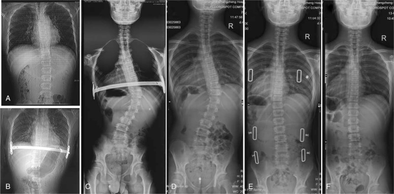
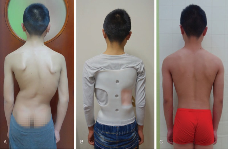
Patient concerns: A 14-year-old boy was referred to pediatric surgery for pectus excavatum deformity. He underwent a successful Nuss procedure. At follow up, the patient was noted having an asymmetric back whole spine X-ray showed a right-sided thoracic curve with a Cobb angle of 54 degrees.
Interventions and outcomes: We obtained a satisfactory result by removing the pectus bar and prescribing the patient a brace.
Lessons: This report demonstrates that the spine should be evaluated routinely before and after Nuss procedure. Besides, spinal fusion is not recommended for acquired scoliosis following pectus excavatum surgery.
Conflict of interest statement
The authors declare no conflict of interest.
Figures
Similar articles
-
Scoliosis Progression After the Nuss Procedure for Pectus Excavatum: A Case Report.Spine Deform. 2019 Nov;7(6):1003-1009. doi: 10.1016/j.jspd.2019.01.009. Spine Deform. 2019. PMID: 31731992
-
Pectus excavatum and scoliosis: a review about the patient's surgical management.Gen Thorac Cardiovasc Surg. 2020 Nov;68(11):1225-1233. doi: 10.1007/s11748-020-01496-y. Epub 2020 Sep 29. Gen Thorac Cardiovasc Surg. 2020. PMID: 32990868 Review.
-
Acquired thoracic scoliosis following minimally invasive repair of pectus excavatum.Am Surg. 2003 Jun;69(6):530-3. Am Surg. 2003. PMID: 12852514
-
Scoliosis after pectus excavatum correction: does it improve or worsen?Eur J Cardiothorac Surg. 2017 Jul 1;52(1):76-82. doi: 10.1093/ejcts/ezx041. Eur J Cardiothorac Surg. 2017. PMID: 28329150
-
Complications associated with the minimally invasive repair of pectus excavatum.Semin Pediatr Surg. 2018 Jun;27(3):151-155. doi: 10.1053/j.sempedsurg.2018.05.001. Semin Pediatr Surg. 2018. PMID: 30078485 Review.
Cited by
-
Clinical significance of concomitant pectus deformity and adolescent idiopathic scoliosis: systematic review with best evidence synthesis.N Am Spine Soc J. 2022 Jun 25;11:100140. doi: 10.1016/j.xnsj.2022.100140. eCollection 2022 Sep. N Am Spine Soc J. 2022. PMID: 35814492 Free PMC article. Review.
-
Impact of the Nuss procedure on spinal curvature across four time points: a longitudinal and subgroup analysis.J Thorac Dis. 2025 Jul 31;17(7):4633-4643. doi: 10.21037/jtd-2025-357. Epub 2025 Jul 21. J Thorac Dis. 2025. PMID: 40809263 Free PMC article.
References
-
- Park HJ, Jeong JY, Jo WM, et al. Minimally invasive repair of pectus excavatum: a novel morphology-tailored, patient-specific approach. J Thorac Cardiovasc Surg 2010;139:379–86. - PubMed
-
- Felts E, Jouve JL, Blondel B, et al. Child pectus excavatum: correction by minimally invasive surgery. Orthop Traumatol Surg Res 2009;95:190–5. - PubMed
-
- Niedbala A, Adams M, Boswell WC, et al. Acquired thoracic scoliosis following minimally invasive repair of pectus excavatum. Am Surg 2003;69:530–3. - PubMed
-
- Nuss D, Kelly RE, Jr, Croitoru DP, et al. A 10-year review of a minimally invasive technique for the correction of pectus excavatum. J Pediatr Surg 1998;33:545–52. - PubMed
Publication types
MeSH terms
LinkOut - more resources
Full Text Sources
Medical
