. 2019 May 8;119(9):5537-5606.
doi: 10.1021/acs.chemrev.8b00532. Epub 2019 Jan 4.

Folding and Misfolding of Human Membrane Proteins in Health and Disease: From Single Molecules to Cellular Proteostasis

Affiliations

Folding and Misfolding of Human Membrane Proteins in Health and Disease: From Single Molecules to Cellular Proteostasis

Justin T Marinko et al. Chem Rev. .

Abstract

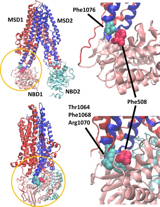
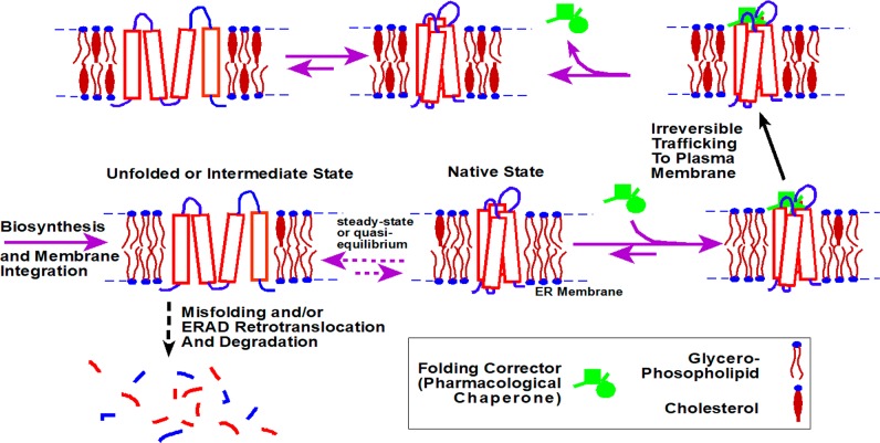
Advances over the past 25 years have revealed much about how the structural properties of membranes and associated proteins are linked to the thermodynamics and kinetics of membrane protein (MP) folding. At the same time biochemical progress has outlined how cellular proteostasis networks mediate MP folding and manage misfolding in the cell. When combined with results from genomic sequencing, these studies have established paradigms for how MP folding and misfolding are linked to the molecular etiologies of a variety of diseases. This emerging framework has paved the way for the development of a new class of small molecule "pharmacological chaperones" that bind to and stabilize misfolded MP variants, some of which are now in clinical use. In this review, we comprehensively outline current perspectives on the folding and misfolding of integral MPs as well as the mechanisms of cellular MP quality control. Based on these perspectives, we highlight new opportunities for innovations that bridge our molecular understanding of the energetics of MP folding with the nuanced complexity of biological systems. Given the many linkages between MP misfolding and human disease, we also examine some of the exciting opportunities to leverage these advances to address emerging challenges in the development of therapeutics and precision medicine.


Conflict of interest statement

The authors declare no competing financial interest.

Figures

Figure 1
Figure 1
Depth-dependent statistical distributions of amino acids within transmembrane domains. Experimental depth dependent compositional biases within transmembrane domains were used to train a probabilistic potential energy function (A) charged, (B) hydrophobic, (C) polar, and (D) aromatic amino acid. Lower energies correspond to higher probabilities of finding the corresponding residue at a given depth. An X-coordinate of 0 Å corresponds to the center of the membrane normal. Reprinted with permission from ref (44). Copyright 2005 John Wiley and Sons.
Figure 2
Figure 2
Adaptation of transmembrane domains to variations in bilayer thickness. Cartoons depict ways TM domains adapt to changes in bilayer thickness. A thinning of the membrane may cause TM domains to tilt with respect to the membrane (top). Alternatively, the bilayer may be locally distorted in order to facilitate the solvation of a lengthy TM domain (middle). Thickening of the bilayer may also result in the extension of TM helices out of the bilayer when sites located at the end of the helix have similar preferences for aqueous exposure or membrane burial (bottom).
Figure 3
Figure 3
Lipid compositions of membranes from various organelles in mammalian cells. Lipid compositions of organelle membranes from rat liver cells (derived from multiple sources) are shown. Abbreviations: PC: phosphatidylcholine, PE: phosphatidylethanolamine, SM: sphingomyelin, PI: phosphatidylinositol, Chol: cholesterol. The data on lipid composition is from ref (92).
Figure 4
Figure 4
Comparison of MP structures from the disparate domains of life. (A) Superpositions of structures of thermophilic archaeal MPs on those of mesophilic counterparts reveal high similarity. Superposition of porins TtoA from Thermus thermophilus (green, PDB ID: 3DZM) and OmpA from E. coli (magenta, PDB ID: 1QJP). (Middle panel) Superposition of ammonium transporters Amt-1 from the archaeal hyperthermophile A. fulgidus (green, PDB ID: 2B2H) and AmtB from E. coli (magenta, PDB ID: 1U77). (Bottom panel) Superposition of aquaporin from A. fulgidus (green, PDB ID: 3NE2) with AqpZ from E. coli (magenta, PDB ID: 1RC2). View onto the membrane surface is from the periplasm/extracellular space. (B) The lipid-contact faces of aquaporins from three domains of life exhibit common features. Shown are aquaporins from a hyperthermophilic archaebacterium, A. fulgidus (PDB ID: 3NE2, left), E. coli (PDB ID: 1RC2, center), and O. airies (sheep) (PDB ID: 3M9I, right). Residues are colored as follows; red: polar residues; blue: large hydrophobic; green: aromatic/His; purple: small (Gly, Ala, Ser, Cys). Figure used with permission from ref (172). Copyright 2015 ACS.
Figure 5
Figure 5
Folding equilibrium for a MP in lipid bilayers versus in detergent micelles. The native conformation is often the most favorable conformational state for MPs in both bilayers and micelles. However, the structural properties of the accessible denatured states, the energies between folded and unfolded states, and the magnitude of the energetic barriers that separate them from the native state may often differ in bilayers and in micelles.
Figure 6
Figure 6
Folding equilibrium for WT and L16P mutant PMP22. This is determined using NMR and other methods under conditions where PMP22 is solubilized in tetradecylphosphocholine micelles at 25 °C. The L16P disease mutation site is located in the first TM helix with the proline substitution resulting in the flexible hinge illustrated in the lower left panel. We suggest that the “partially unfolded state” depicted on the left may actually be similar to the true physiological unfolded state. Further unfolding is restrained by the short loops connect TM2 to TM3 and TM3 to TM4. Reprinted with permission from ref (182). Copyright 2011 Cell Press.
Figure 7
Figure 7
Classes of model membrane used in studies of purified MPs. Not illustrated here are “lipodisqs”, which resemble nanodiscs except that a synthetic amphipathic polymer is used to stabilize the edge of the bilayered disc instead of an amphipathic protein.
Figure 8
Figure 8
Effect of ligand binding on the kinetic stability of integral membrane proteins. If the native binding pocket is disrupted prior to the rate limiting step for unfolding, then excess ligand will selectively stabilize the native conformation (N) relative to the transition state for unfolding (‡) and the denatured state (red). In this case, the rate of unfolding and the fraction of unfolded protein at equilibrium will be decreased in the presence of ligand.
Figure 9
Figure 9
Hypothetical morphology of the conformational energy landscape for bR in DMPC/CHAPSO/SDS bicelles. This cartoon depicts a hypothetical energy landscape that describe the conformational energetics of bR in bicelles. The upper rim of the conformational energy landscape represents the random coil state, which is unlikely to be sampled within membranous environments. Instead, the TM segments are likely to persist in an ensemble of helical bundles within the denatured state, which is represented by the secondary basin of the energy landscape. To find the native conformation, helical TM segments must explore various topological configuration until the native topology is achieved and folding can proceed downhill. It is emphasized that, for many membrane proteins, the folding funnel may be much more complicated than as proposed here for the well-characterized case of BR.
Figure 10
Figure 10
Single-molecule forced unfolding of a MP, GlpG. (A) Schematic of the single-molecule magnetic tweezers experiment for studying the unfolding and refolding of GlpG in a bicelle. The protein termini have been conjugated with DNA, with the end of one DNA molecule being surface anchored and the other end being attached to a bead that can be pulled away from the surface to force unfolding of the protein in the plane of the bicelle bilayer. (B) Representative force–extension curves for repeated GlpG unfolding and refolding transitions. (C) The energy landscape for folding/unfolding of GlpG in bicelles, where kf0 and ku0 are the kinetic rates for folding and unfolding at zero force, ΔG is the unfolding free energy, and ΔGu† and ΔGf† are the kinetic energy barriers for unfolding and folding, respectively. Reprinted with adaptations from ref (269). Copyright 2015 Springer Nature.
Figure 11
Figure 11
Assay to measure the efficiency of spontaneous insertion, folding, and trimerization of DAGK into preformed lipid vesicles following a many-fold dilution of a small aliquot of the pure enzyme in lipid and detergent-free urea or guanidinium solutions. Successful folding of the protein is accompanied by the appearance of enzyme activity that is monitored through a spectrophotometrically detected coupled assay system. The degree of misfolding is assessed based on comparing the final observed enzyme activity with the activity expected for 100% folding efficiency. Abbreviations: DAG, diacylglycerol; PEP, phosphenolpyruvate; NAD+ and NADH, oxidized and reduced forms of nicotinamidedinucleotide. Figure used with permission from ref (192). Copyright 2015 ACS.
Figure 12
Figure 12
(A) Structure of the ribosome-translocon complex. A 3.4 Å resolution model of a mammalian ribosome (blue and brown) bound to the Sec61 complex (red) determined by single particle Cryo-EM is shown. The absence of tRNA in the peptidyl transfer center (PTC) indicates this complex represents the inactive conformation of the complex. Subtle structural rearrangements have been observed within the active complex (not shown). Panel adapted with permission from (359). Copyright 2014, Elsevier under CC BY 3.0 https://creativecommons.org/licenses/by/3.0/. (B) Sec61 translocon and complex with the ribosome, TRAP, and the oligosaccharyl transferase. Segmented densities for the 40S (yellow) and 60S (light blue) ribosomal subunits, translation elongation factors (magenta), Sec61 (blue), TRAP (green), and OST (red) from a subtomogram average of the ER membrane associated translocon complex filtered to 9.0 Å resolution. Figure adapted with permission from ref (365). Copyright 2015 Nature under CC BY 4.0 http://creativecommons.org/licenses/by/4.0/.
Figure 13
Figure 13
Overview of MP folding in the early secretory pathway of mammalian cells. This figure encompasses ERAD, ERAF, and ERES, plus some components of the broader proteostatic network.
Figure 14
Figure 14
N-Linked glycan (A) and the calnexin cycle (B). As proteins are translocated into the ER membrane they are tagged with a 14-sugar moiety at N-X-S/T (where X is any amino acid but P) sequence motifs as shown in panel A. This post-translational modification serves a multitude of functions for the protein including increasing hydrophilicity, preventing aggregation, influencing tertiary contacts, and serving as a folding “barcode”. Through sequential cleavage of monosaccharides (gray dotted lines), the folding polypeptide can be engaged with different lectin chaperones involved in either ERAF or ERAD. The enzymes responsible for cleaving various glyosidic linkages are shown. The predominant folding pathway for glycosylated proteins is shown in panel B. Monoglucosylated proteins (1) are engaged by the membrane-anchored lectin chaperone calnexin (2, gray). Calnexin provides the protein with an isolated environment to fold, recruits essential cochaperones such as the disulfide isomerase ERp57 (cyan), and may also facilitate the proper tertiary packing of a MP by associating with exposed hydrophilic residues in the plane of the membrane. Association is transient; glucosidase II cleaves the terminal glucose residue on the glycan chain freeing the client polypeptide from engagement with calnexin. Proteins that have yet to attain their proper tertiary structure (3), are sensed by UGGT1 (orange) and reglucosylated, allowing reentry into the calnexin cycle (1). Proteins that have obtained their proper structure and post-translational modifications (particularly disulfide bonding) are sensed by UGGT1 and funneled toward the ER export machinery for ER exit (4). Polypeptides that fail to complete folding after consecutive cycles are eventually funneled out of the cycle and targeted for ERAD by the action of mannosidases.
Figure 15
Figure 15
Some of the possible folding defects in MPs that must be recognized and managed by the folding quality control systems of all cells.
Figure 16
Figure 16
Examples of mechanisms for ERQC detection of a misfolded integral MP. In panel A, the 10 TM α subunit (red) of the Na+, K+ ATPase is able to properly integrate into the ER membrane in the presence of the single-pass TM β subunit (blue). In the absence of the β subunit, TM helices 7 and 8 fail to properly integrate into the ER membrane and sidle into the cytoplasm where they can be recognized by the Hsp70 chaperone (black), leading to targeting for degradation. In panel B, a hypothetical dual pass TM protein (cyan) requires a salt-bridge (acidic and basic amino acids shown with stars) in the plane of the membrane in order to maintain its proper fold. If the protein is misfolded or unstable, it can expose these hydrophilic amino acids in the plane of the membrane. Hrd1 (green) contains a number of hydrophilic amino acids in the plane of the membrane that may aid in recognizing aberrantly exposed polar residues in MPs, resulting in ERAD targeting.
Figure 17
Figure 17
ERAD of a representative integral MP (red). In step 1, a misfolded protein (red) is recognized either through its N-glycan by the ER lectin chaperone OS9 (orange) or in the plane of the membrane by proteins such as the derlins (gray) or Hrd1 (green). Sel1L (purple) nucleates an ERAD complex in the ER membrane and also recruits ER luminal factors such as BiP and its PDI cochaperone ERdJ5 (teal). Herp (cyan) localizes the E3 ubiquitin ligase Hrd1 to ERAD sites. In step 2, the derlins may function to lower the energetic barrier for substrate retrotranslocation by partially unwinding helices in the plane of the membrane. The cytoplasmic region of Hrd1 catalyzes the addition of ubiquitin (brown) to lysine residues on the ERAD substrate. Ufd1 and NPL4 (dark orange) associate with the AAA ATPase p97 (yellow) and recognize the ubiquitinated ERAD substrate. In step 3, the ERAD substrate is retrotranslocated into the cytoplasm potentially through a pore formed by Hrd1. PNGase (light green) then removes the N-linked glycan from the ERAD substrate. The substrate is pulled out of the membrane via the energy provided by p97 ATPase activity which also functions as a retrochaperone to maintain the solubility of the ERAD substrate in the cytoplasm. The ERAD substrate is eventually degraded via the 26S proteasome (gray and navy).
Figure 18
Figure 18
To-scale surface representation the structures of some of the key players involved in ERAD. Human BiP (black; PDB: 5E84), Hrd1 from S. cerevisiae (green; PDB: 5 V6P), human p97 (yellow; PDB: 5FTJ), and the human 20S proteasome (α subunits in gray, β subunits in navy; PDB: 5LE5).
Figure 19
Figure 19
Documented nephrogenic diabetes insipidus mutations in the vasopressin V2 receptor. List of mutation is from.
Figure 20
Figure 20
(A) Growth with time in the total number of identified human mutations that result in inherited (Mendelian) monogenic disorders Data from the Human Gene Mutation Database. (B) Growth with time in the total number of validated human genome variations (SNVs and other small scale variations) as logged in the online dbSNP Database. The small decreases in the number of variations seen in this plot for some time points reflect the consequences of changes in the reference genome and its annotation with time. Figure adapted with permission from ref (629). Copyright 2015 ACS.
Figure 21
Figure 21
Structure of KCNQ1 and location of mutations examined in Huang et al. (2018). (Left) Cryo-EM structure of Xenopus KCNQ1, highlighting one of its four voltage sensor domains. Note that some of the connecting loops between TM helices were not resolved in the EM structure and are therefore not depicted. (Center and Right) Orthogonal views of an isolated voltage sensor domain with side chains shown for sites that correspond to those experimentally characterized by Huang et al. in their study of the human KCNQ1 channel. Blue sites are those where mutations did not dramatically alter the stability or trafficking of KCNQ1. These sites are seen to be enriched on the surface of the domain. Red sites correspond to mutants that were seen to cause both mistrafficking in cells and (usually) instability of the voltage sensor domain under NMR conditions. These sites tend to cluster in the interior of the VSD.
Figure 22
Figure 22
Results from characterizing the channel function, trafficking, and stability of 51 mutant forms of the human KCNQ1 potassium channel. (A) KCNQ1 potassium channel cell surface expression levels versus measured K+ channel peak current density as determined in HEK293 cells. Data are color-coded: known LQTS disease mutants (red), variants of unknown significance (VUS) observed in humans but not previously classified (cyan), or predicted neutral polymorphism (black). The vertical red lines indicate values that are 65% of WT, corresponding to the approximate cutoff between “healthy” and LQTS-predisposing. These data illustrate that for a majority of disease mutants, loss of channel function is the consequence of failure of the channel to traffic to the cell surface. It is also seen that a number of the VUS mutants exhibit loss of channel function, again usually as a result of mistrafficking. These VUS are likely LQTS-predisposing. (B) 1H,15N-TROSY NMR spectrum of the WT KCNQ1 voltage sensor domain (residues 100 to 249) and (C) spectrum of a mistrafficking-prone disease mutant form of KCNQ1, E115G (red) superimposed on the WT spectrum (back). These spectra were acquired for the two forms of the VSD solubilized in lyso-myristoylphosphatidylglycerol (LMPG) micelles. The spectrum of the E115G disease mutant exhibits extensive broadening, disappearance, and shifts of peaks, indicating that its structure is destabilized relative to that of the WT protein. NMR revealed that the vast majority of the mistrafficking-prone mutants were folding-destabilized. Adapted with permission from (183). Copyright 2018 American Association for the Advancement of Science.
Figure 23
Figure 23
Four views of the structure of rhodopsin, illustrating the sites of known retinitis pigmentosa point mutations (green). The retinal cofactor is violet. PDB code: 1L9H.
Figure 24
Figure 24
Thermodynamic destabilization of human PMP22 results in mistrafficking of the protein and Charcot-Marie Tooth disease (peripheral neuropathy). Panels A and B show the locations of the disease mutants in the sequence and modeled 3-D structure of the protein, respectively. Panel C shows a strong correlation between surface trafficking efficiency and stability across the panel of tested PMP22 mutants. Panel D shows that the extent of surface trafficking correlates well with nerve conduction velocity in humans carrying each mutant form. Healthy patients present with high conduction velocities, with reductions in conduction velocity correlating with disease severity. Panel E shows that there is also a strong correlation between the stability of PMP22 and nerve conduction velocity. Figures adapted with permission from ref (287). Copyright 2015 ACS.
Figure 25
Figure 25
Structures of CFTR in its resting conformation (top) and in its phosphorylated+ATP-bound state (bottom). The side chain of Phe508 shown in van der Waals form (red) along with the interacting residues (cyan) from the hairpin connecting the 10th and 11th TM helices (blue). PDB: 5TSI (top) and 6MSM (bottom).
Figure 26
Figure 26
Hypothetical scenario of action for a PC as a drug. In this case the PC selectively binds to and stabilizes the folded form of the with target MP, tipping the balance between correct folding/trafficking and ERAD degradation in favor of folding and trafficking. The initial binding/rescue event occurs shortly after administration of the PC as a drug, at which point the PC concentration is fairly high. Once the MP reaches the plasma membrane and the total PC concentration is cleared (due to cytochrome P450 action, for example) the PC will dissociate and not be replenished, at which point the protein remains mostly folded because it is thermodynamically stable in the plasma membrane.
Figure 27
Figure 27
Structures of PCs for the V2 vasopressin receptor. The apparent affinities of each ligand for the WT V2R are indicated.
Figure 28
Figure 28
Structures of PCs for the gonadotropin-releasing hormone receptor. The apparent affinities of each ligand for the WT GnRHR are indicated.,
Figure 29
Figure 29
Structures of PCs for the melanocortin-4 receptor. The apparent ligand affinities for the WT MC4R are indicated.
Figure 30
Figure 30
Structures of functional potentiators (A) and PCs (B) for the CFTR channel. The apparent ligand affinities or rescue potencies of each compound for the ΔF508 or G551D mutant forms of CFTR are indicated.,− RDR1 rescues ΔF508 CFTR at micromolar concentrations.
Figure 31
Figure 31
Examples of cell-based HTS for pharmacological chaperones that restore plasma membrane trafficking (A) or function (B) of a MP, in this case a GPCR. Cells stably expressing a misfolded mutant of the target MP are treated with different compounds (represented as small colored balls). Compounds that act as PCs stabilize the misfolded protein and facilitate its trafficking to the plasma membrane where its expression and/or function can be detected or reported.

Similar articles

Cited by

References

    1. Deisenhofer J.; Epp O.; Miki K.; Huber R.; Michel H. Structure of the protein subunits in the photosynthetic reaction centre of Rhodopseudomonas viridis at 3A resolution. Nature 1985, 318, 618–624. 10.1038/318618a0. - DOI - PubMed
    1. Kendrew J. C.; Bodo G.; Dintzis H. M.; Parrish R. G.; Wyckoff H.; Phillips D. C. A three-dimensional model of the myoglobin molecule obtained by x-ray analysis. Nature 1958, 181, 662–666. 10.1038/181662a0. - DOI - PubMed
    1. Huang K. S.; Bayley H.; Liao M. J.; London E.; Khorana H. G. Refolding of an integral membrane protein. Denaturation, renaturation, and reconstitution of intact bacteriorhodopsin and two proteolytic fragments. J. Biol. Chem. 1981, 256, 3802–3809. - PubMed
    1. London E.; Khorana H. G. Denaturation and renaturation of bacteriorhodopsin in detergents and lipid-detergent mixtures. J. Biol. Chem. 1982, 257, 7003–7011. - PubMed
    1. Popot J. L.; Gerchman S. E.; Engelman D. M. Refolding of bacteriorhodopsin in lipid bilayers. A thermodynamically controlled two-stage process. J. Mol. Biol. 1987, 198, 655–676. 10.1016/0022-2836(87)90208-7. - DOI - PubMed

Publication types

Substances