. 2019 Mar 1;18(3):947-959.
doi: 10.1021/acs.jproteome.8b00647. Epub 2019 Jan 23.

Quantitative Proteomic Analysis of Small and Large Extracellular Vesicles (EVs) Reveals Enrichment of Adhesion Proteins in Small EVs

Affiliations

Quantitative Proteomic Analysis of Small and Large Extracellular Vesicles (EVs) Reveals Enrichment of Adhesion Proteins in Small EVs

Lizandra Jimenez et al. J Proteome Res. .

Abstract

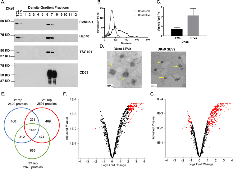
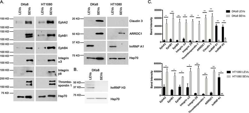
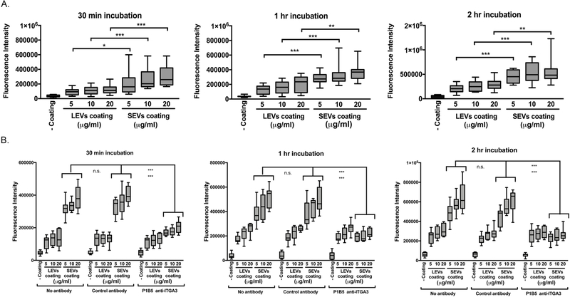
Extracellular vesicles (EVs) are important mediators of cell-cell communication due to their cargo content of proteins, lipids, and RNAs. We previously reported that small EVs (SEVs) called exosomes promote directed and random cell motility, invasion, and serum-independent growth. In contrast, larger EVs (LEVs) were not active in those assays, but might have unique functional properties. In order to identify protein cargos that may contribute to different functions of SEVs and LEVs, we used isobaric tags for relative and absolute quantitation (iTRAQ)-liquid chromatography (LC) tandem mass spectrometry (MS) on EVs isolated from a colon cancer cell line. Bioinformatics analyses revealed that SEVs are enriched in proteins associated with cell-cell junctions, cell-matrix adhesion, exosome biogenesis machinery, and various signaling pathways. In contrast, LEVs are enriched in proteins associated with ribosome and RNA biogenesis, processing, and metabolism. Western blot analysis of EVs purified from two different cancer cell types confirmed the enrichment of cell-matrix and cell-cell adhesion proteins in SEVs. Consistent with those data, we found that cells exhibit enhanced adhesion to surfaces coated with SEVs compared to an equal protein concentration of LEVs. These data suggest that a major function of SEVs is to promote cellular adhesion.

Keywords: adhesion; exosomes; extracellular vesicles; iTRAQ; microvesicles; proteomics.


Conflict of interest statement

Conflict of interest statement

No potential conflicts of interest were disclosed.

Figures

Figure 1.
Figure 1.. Characterization and proteomic analysis of DKs8 EVs.
A. Western blot analysis of DKs8 total cell lysate, LEVs and density gradient fractions for Flotillin, Hsp70, Tsg101 and CD63. B. Representative traces from nanoparticle tracking analysis of DKs8 LEVs and SEVs. C. Quantitation of vesicle numbers from nanoparticle tracking analysis (n=3). D. Representative TEM images of DKs8 LEVs and SEVs. Yellow arrows point to the EVs. Scale bar shows 100 nm. E. Venn diagram showing the overlap of the proteins identified in the three iTRAQ replicates. F. Volcano plot highlighting proteins having p<0.01 and absolute log2 fold change >2 in red. G. Volcano plot highlighting proteins having p<0.05 and absolute log2 fold change >1 in red.
Figure 2.
Figure 2.. Western blot validation of selected proteins in both the DKs8 and HT1080 EVs.
A. Western blot analysis of DKs8 and HT1080 EVs validating selected proteins (EphA2, EphB1, EphB4, Integrin α3, Integrin β8, Thrombospondin-1, ARRDC1, Claudin 3, hnRNP A1) identified in the iTRAQ study. B. Western blot analysis of DKs8 EVs validating hnRNP H3 identified in the iTRAQ study. C. Quantitation of Western blots from ≥3 independent experiments. * p < 0.05; ** p < 0.01; *** p < 0.001 comparing the band intensities of the DKs8 or HT1080 LEVs and SEVs.
Figure 3.
Figure 3.. Western blot analysis confirmed cell adhesion proteins are still present in ARRDC1 KD SEVs.
A. Western blot analysis of DKs8 shScramb. and shARRDC1-KD TCLs for ARRDC1 and Beta actin. B. Representative nanoparticle tracking traces of SEVs from DKs8 shScramb. and shARRDC1-KD cells. C. Quantitation of SEVs numbers from DKs8 shScramb. and shARRDC1–2 and −3 cells determined in nanoparticle tracking analysis (n=4). D. Western blot analysis of DKs8 shScramb. and shARRDC1-KD SEVs assessing the levels of EphA2, EphB1, EphB4, Integrin α3, Integrin β8, Thrombospondin-1, ARRDC1, Claudin 3 and Hsp70. E. Quantitation of Western blots from ≥3 independent experiments. * p < 0.05; *** p < 0.001 comparing the band intensities of DKs8 shScramb., shARRDC1-KD SEVs.
Figure 4.
Figure 4.. Small EVs promoted cell adhesion of DKs8 cells.
A. Calcein AM fluorescence of adherent cells was measured. In each of the independent experiments, all of conditions were performed in triplicate. Data is shown from four independent experiments. Note that at all three time points and for all three coating concentrations, both the LEVs and SEVs significantly promoted DKs8 adhesion compared to the no coating condition (p < 0.001). * p < 0.05; ** p < 0.01; *** p < 0.001 compared to equal LEVs and SEVs coating concentrations. B. Calcein AM fluorescence of adherent cells was measured for the cell adhesion assay in the presence of an integrin α3 blocking antibody (P1B5), a matched IgG control antibody or no antibody. In each of the independent experiments, all of conditions were performed in triplicate. Data is shown from three independent experiments. Note that the enhancements in cell adhesion by SEVs in the matched IgG conditions were not significantly altered when you compare the SEV-no antibody conditions at all three time points and for all three coating concentrations. *** p < 0.001 for equal SEVs coating concentrations in the presence of no antibody (top ***) and matched IgG (bottom ***) compared to in the presence of P1B5 integrin α3 blocking antibody.

References

    1. Maas SLN; Breakefield XO; Weaver AM, Extracellular Vesicles: Unique Intercellular Delivery Vehicles. Trends Cell Biol 2017, 27, (3), 172–188. - PMC - PubMed
    1. van Niel G; D’Angelo G; Raposo G, Shedding light on the cell biology of extracellular vesicles. Nature Reviews Molecular Cell Biology 2018, 19, (4), 213–228. - PubMed
    1. Kowal J; Arras G; Colombo M; Jouve M; Morath JP; Primdal-Bengtson B; Dingli F; Loew D; Tkach M; Thery C, Proteomic comparison defines novel markers to characterize heterogeneous populations of extracellular vesicle subtypes. Proc Natl Acad Sci U S A 2016, 113, (8), E968–77. - PMC - PubMed
    1. Zhang H; Freitas D; Kim HS; Fabijanic K; Li Z; Chen H; Mark MT; Molina H; Martin AB; Bojmar L; Fang J; Rampersaud S; Hoshino A; Matei I; Kenific CM; Nakajima M; Mutvei AP; Sansone P; Buehring W; Wang H; Jimenez JP; Cohen-Gould L; Paknejad N; Brendel M; Manova-Todorova K; Magalhaes A; Ferreira JA; Osorio H; Silva AM; Massey A; Cubillos-Ruiz JR; Galletti G; Giannakakou P; Cuervo AM; Blenis J; Schwartz R; Brady MS; Peinado H; Bromberg J; Matsui H; Reis CA; Lyden D, Identification of distinct nanoparticles and subsets of extracellular vesicles by asymmetric flow field-flow fractionation. Nat Cell Biol 2018, 20, (3), 332–343. - PMC - PubMed
    1. Willms E; Cabanas C; Mager I; Wood MJA; Vader P, Extracellular Vesicle Heterogeneity: Subpopulations, Isolation Techniques, and Diverse Functions in Cancer Progression. Front Immunol 2018, 9, 738. - PMC - PubMed

Publication types

Substances