. 2019 Jan 4;17(1):e2006926.
doi: 10.1371/journal.pbio.2006926. eCollection 2019 Jan.

Differential and convergent utilization of autophagy components by positive-strand RNA viruses

Affiliations

Differential and convergent utilization of autophagy components by positive-strand RNA viruses

Emma Abernathy et al. PLoS Biol. .

Abstract

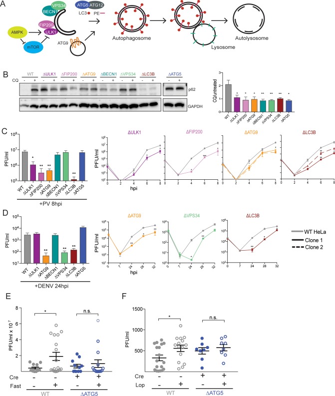
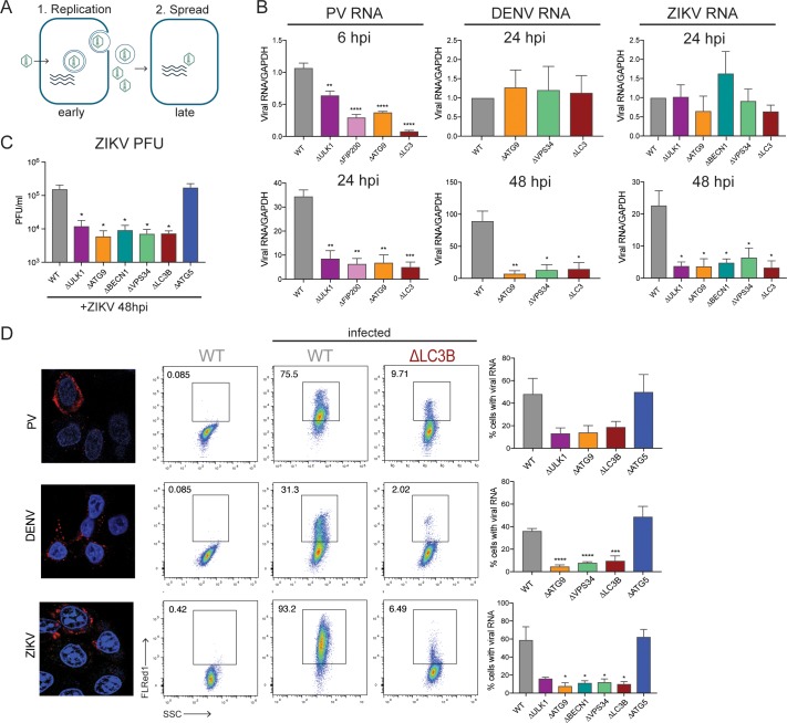
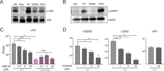
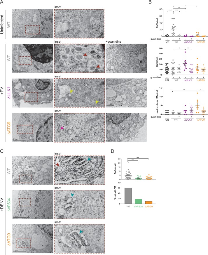
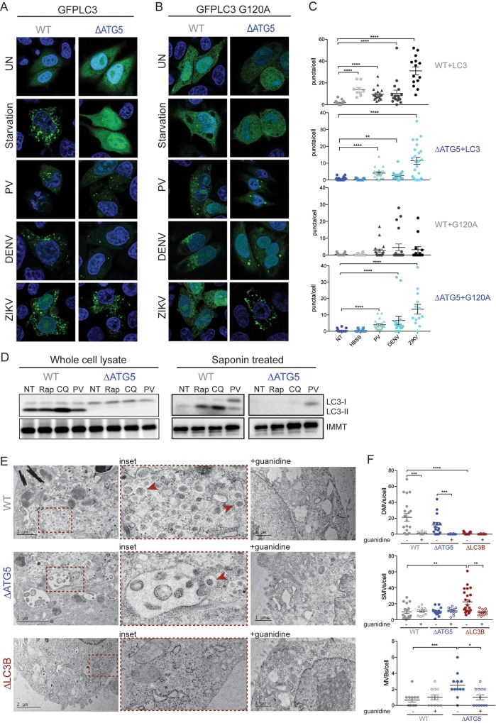
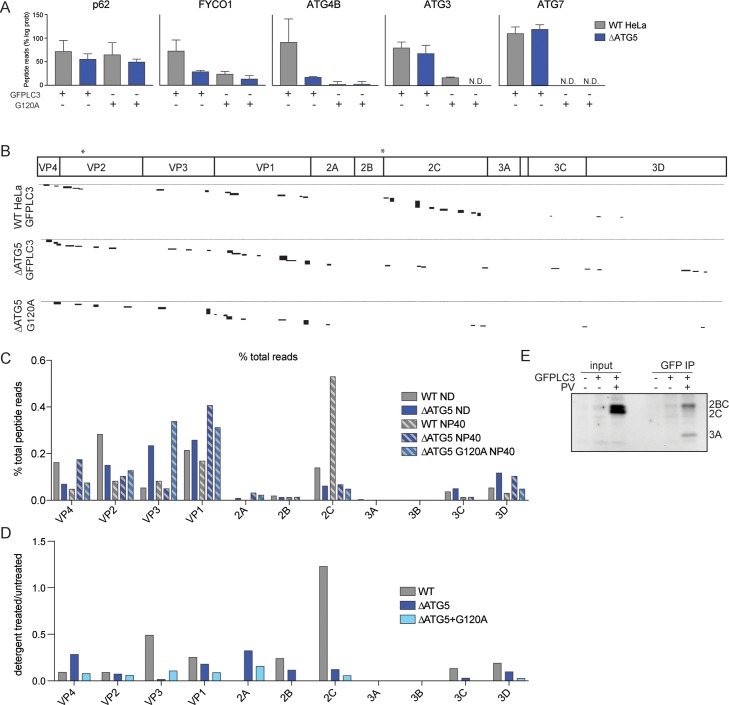
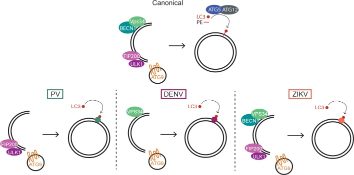
Many viruses interface with the autophagy pathway, a highly conserved process for recycling cellular components. For three viral infections in which autophagy constituents are proviral (poliovirus, dengue, and Zika), we developed a panel of knockouts (KOs) of autophagy-related genes to test which components of the canonical pathway are utilized. We discovered that each virus uses a distinct set of initiation components; however, all three viruses utilize autophagy-related gene 9 (ATG9), a lipid scavenging protein, and LC3 (light-chain 3), which is involved in membrane curvature. These results show that viruses use noncanonical routes for membrane sculpting and LC3 recruitment. By measuring viral RNA abundance, we also found that poliovirus utilizes these autophagy components for intracellular growth, while dengue and Zika virus only use autophagy components for post-RNA replication processes. Comparing how RNA viruses manipulate the autophagy pathway reveals new noncanonical autophagy routes, explains the exacerbation of disease by starvation, and uncovers common targets for antiviral drugs.


Conflict of interest statement

The authors have declared that no competing interests exist.

Figures

Fig 1
Fig 1. Diverse viruses utilize different components of the autophagy pathway.
(A) Schematic of the autophagy pathway and the components targeted in this study for KO by CRISPR-Cas9. (B) Human HeLa cells were left untreated or treated with CQ for 2 hours. Protein lysates were collected and immunoblotted for p62 and GAPDH. Quantification of autophagic flux was done by comparing p62/GAPDH in CQ and untreated samples. (C and D) HeLa cells were infected with PV or DENV at a MOI of 0.1; PFU/cell and cells (PV) or supernatant (DENV) were harvested at indicated times post infection for titration by plaque assay. (E and F) C57BL/6 PVR+/+ Atg5fl/fl Cre+/− mice were fasted for 48 hours and then infected intramuscularly with PV for E or administered loperamide every 12 hours by i.p. injection during the 4 days of infection for F. Calf muscle tissue was collected and titered by plaque assay at 4 dpi. Open circles denote fasted or drug-treated mice and blue circles indicate Atg5-deficient mice. All data are represented as mean +/− SEM. *Indicates significant P value of <0.05, **P value < 0.01, ***P value < 0.001, ****P value > 0.0001 by an unpaired t test (C and D) or a Mann–Whitney test (E–F). See also S1 and S2 Figs, S1 and S2 Tables, and S1 Data. Atg5, autophagy-related gene 5; CRISPR, Clustered Regularly Interspaced Short Palindromic Repeats; CQ, chloroquine; DENV, dengue virus; dpi; GAPDH, glyceraldehyde 3-phosphate dehydrogenase; HeLa, human epithelial-derived cell line; i.p., intraperitoneal; KO, knockout; MOI, multiplicity of infection; PFU, plaque-forming units; PV, poliovirus; PVR, poliovirus receptor.
Fig 2
Fig 2. PV uses components of the autophagy pathway for early replicative events, while DENV and ZIKV use it for post-RNA replication processes.
(A) Schematic of viral replication and spread. (B) HeLa cells were infected with PV, DENV, or ZIKV at an MOI of 0.1 PFU/cell, and RNA was harvested at 6, 24, or 48 hpi. Viral RNA abundance was measured by RT-qPCR and normalized to GAPDH. (C) HeLa cells were infected with ZIKV at an MOI of 0.1 PFU/cell, and supernatant was harvested at 48 hpi. Viral titers were determined by plaque assay. (D) WT HeLa cells were infected with PV, DENV, or ZIKV at an MOI of 1 PFU/cell for 24 hours. Cells were fixed and stained with viral-specific fluorescent probes and visualized by confocal microscopy. For flow cytometry, HeLa cells were infected at an MOI of 0.1 PFU/cell for 24 hours (PV) or 48 hours (DENV, ZIKV). Cells were fixed and stained with viral-specific probes. Percent infection was determined by gating on the uninfected controls. Representative FACS plots are shown. All data are represented as mean +/− SEM. *Indicates significant P value of <0.05, **P value < 0.01, ***P value < 0.001, ****P value > 0.0001 by an unpaired t test. See also S3 Fig and S2 Data. DENV, dengue virus; GAPDH, glyceraldehyde 3-phosphate dehydrogenase; HeLa, human epithelial-derived cell line; hpi, hours post infection; MOI, multiplicity of infection; PFU, plaque-forming units; PV, poliovirus; RT-qPCR, reverse transcription quantitative PCR; WT, wild-type; ZIKV, Zika virus.
Fig 3
Fig 3. Differential utilization of upstream autophagy components by RNA viruses.
(A and B) Cells were treated with rapamycin or infected with PV (6 hpi), DENV (24 hpi), or ZIKV (24 hpi). Cell lysates were harvested and immunoblotted with phospho-S6K or phospho-AMPK antibodies. (C) Cells were treated with 0.5, 5, or 50 μM of MBL56 and infected with PV at an MOI of 0.1 PFU/cell for 6 hours. (D) WT cells were pretreated with 1, 5, or 10 μM of SAR405 and infected with DENV or ZIKV at an MOI of 0.1 PFU/cell for 24 hours (DENV) or 48 hours (ZIKV). WT cells were pretreated with 10 μM of SAR405 and infected with PV at an MOI of 0.1 PFU/cell for 6 hours. All data are represented as mean +/− SEM. *Indicates significant P value of <0.05, **P value < 0.01, ***P value < 0.001, ****P value > 0.0001 by an unpaired t test. See also S3 Data. AMPK, AMP-activated protein kinase; DENV, dengue virus; hpi, hours post infection; MOI, multiplicity of infection; PFU, plaque-forming units; PV, poliovirus; WT, wild-type; ZIKV, Zika virus.
Fig 4
Fig 4. Virally induced membrane rearrangements are altered in cells that lack individual autophagy components.
(A and B) HeLa cells were infected with PV or DENV at an MOI of 10 PFU/cell. Indicated cells were treated with 2 mM guanidine during the infection period. Cells were fixed at 6 hpi (PV) or 24 hpi (DENV) and subjected to high-pressure freezing and freeze substitution. Images were collected on a TEM microscope. Representative images are shown. Red arrowheads indicate DMVs, yellow arrowheads indicate SMVs, pink arrowheads indicate electron-dense SMVs, blue arrowheads indicate CM. Quantification of cellular structures was done on blinded images, and >10 cells per condition were counted. All data are represented as mean +/− SEM. *Indicates significant P value of <0.05, **P value < 0.01, ***P value < 0.001, ****P value > 0.0001 by a Mann–Whitney test. See also S4 Data. CM, convoluted membranes; DENV, dengue virus; DMV, double-membraned vesicles; HeLa, human epithelial-derived cell line; hpi, hours post infection; MOI, multiplicity of infection; PFU, plaque-forming units; PV, poliovirus; SMV, single-membraned vesicles.
Fig 5
Fig 5. LC3 is recruited to membranes independent of lipidation during viral infection.
(A and B) HeLa cells were transfected with GFP–LC3 or GFP–LC3–G120A for 48 hours. Cells were either starved for 2 hours or infected at an MOI of 10 PFU/cell with PV (6 hours), DENV or ZIKV (24 hours) and fixed for visualization by confocal microscopy. (C) Puncta per cell were counted for each condition; n = >10 cells. (D) HeLa cells were treated with Rap, CQ, or infected with PV at an MOI of 10 PFU/ml for 6 hours. Lysates were harvested with or without saponin and run on an SDS PAGE gel. Immunoblots were stained for LC3 and the membrane-associated IMMT. (E) HeLa cells were infected with PV at an MOI of 10 PFU/cell and subjected to high-pressure freezing and freeze substitution for visualization on a TEM microscope. Representative images are shown. Red arrowheads indicated DMVs. (F) Cell structures were quantified on blinded images; n = >15 cells. All data are represented as mean +/− SEM. *Indicates significant P value of <0.05, **P value < 0.01, ***P value < 0.001, ****P value > 0.0001 by a Mann–Whitney test. See also S5 Data. CQ, chloroquine; DENV, dengue virus; DMV, double-membraned vesicle; GFP, green fluorescent protein; HeLa, human epithelial-derived cell line; IMMT, inner membrane mitochondrial protein; LC3, light-chain 3; MOI, multiplicity of infection; PFU, plaque-forming units; PV, poliovirus; Rap, rapamycin.
Fig 6
Fig 6. Viral proteins bind LC3.
(A) Cells were transfected with GFP–LC3 or GFP–LC3–G120A for 48 hours and infected with PV at an MOI of 10 PFU/cell for 6 hours. An immunoprecipitation was done with anti-GFP antibodies, submitted for mass spectrometry, and peptide read abundance analyzed. Known LC3 interactors were assessed for binding capacity by comparing the log probability of peptide reads to uninfected control samples, which were set to 100%. N.D. indicates no peptide reads were detected. (B) Peptide reads from PV proteins were aligned to the PV genome. Black blocks indicate length and abundance of peptide reads. Stars indicate location of WxxL motifs in the PV genome. (C) PV peptide reads were analyzed by percent total reads for each of the viral proteins. (D) PV peptide reads were analyzed as total reads of detergent-treated samples over nontreated samples. (E) Cells were transfected with GFP–LC3 for 48 hours and infected with PV at an MOI of 10 PFU/cell for 6 hours. Cells were lysed with NP-40 buffer and immunoprecipitated with anti-GFP antibodies. Immunoblots were stained with antibodies against PV proteins 2C and 3A. See also S4 and S5 Figs and S6 Data. GFP, green fluorescent protein; LC3, light-chain 3; MOI, multiplicity of infection; PFU, plaque-forming units; PV, poliovirus.
Fig 7
Fig 7. Model for viral utilization of autophagy pathway components.
Canonical autophagy uses upstream components ULK1/FIP200 complex, VPS34/BECN1 complex, as well as ATG5-mediated lipidation of LC3. PV, DENV, and ZIKV use unique sets of components to induce different membrane structures for viral amplification and spread. ATG5, autophagy-related gene 5; BECN1, beclin-1; DENV, dengue virus; FIP200, PTK2/FAK family interacting protein of 200 kDa; LC3, light-chain 3; PV, poliovirus; ULK1, Unc-like autophagy-activating kinase; ZIKV, Zika virus.

Similar articles

Cited by

References

    1. Mizushima N, Yoshimori T, Ohsumi Y. The role of Atg proteins in autophagosome formation. Annu Rev Cell Dev Biol. 2011;27:107–32. 10.1146/annurev-cellbio-092910-154005 - DOI - PubMed
    1. Tooze SA, Yoshimori T. The origin of the autophagosomal membrane. Nat Cell Biol. 2010;12(9):831–5. 10.1038/ncb0910-831 - DOI - PubMed
    1. Hailey DW, Rambold AS, Satpute-Krishnan P, Mitra K, Sougrat R, Kim PK, et al. Mitochondria supply membranes for autophagosome biogenesis during starvation. Cell. 2010;141(4):656–67. 10.1016/j.cell.2010.04.009 - DOI - PMC - PubMed
    1. Ravikumar B, Moreau K, Jahreiss L, Puri C, Rubinsztein DC. Plasma membrane contributes to the formation of pre-autophagosomal structures. Nat Cell Biol. 2010;12(8):747–57. 10.1038/ncb2078 - DOI - PMC - PubMed
    1. Ge L, Zhang M, Schekman R. Phosphatidylinositol 3-kinase and COPII generate LC3 lipidation vesicles from the ER-Golgi intermediate compartment. Elife. 2014;3:e04135 10.7554/eLife.04135 - DOI - PMC - PubMed

Publication types

MeSH terms