Review
. 2019 Jan 1;8(1):16.
doi: 10.3390/cells8010016.

Relevance of Autophagy in Parenchymal and Non-Parenchymal Liver Cells for Health and Disease

Affiliations
Review

Relevance of Autophagy in Parenchymal and Non-Parenchymal Liver Cells for Health and Disease

Ralf Weiskirchen et al. Cells. .

Abstract

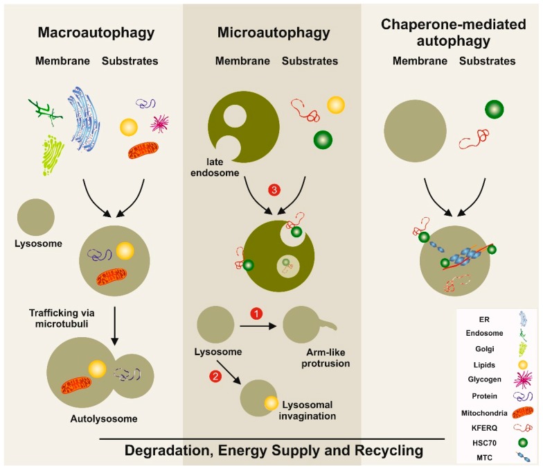
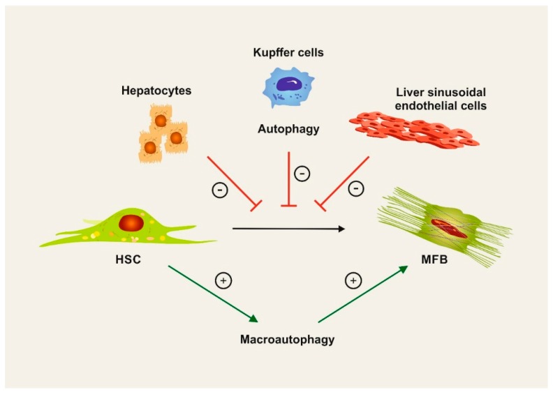
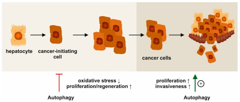
Autophagy is a highly conserved intracellular process for the ordered degradation and recycling of cellular components in lysosomes. In the liver, parenchymal cells (i.e., mainly hepatocytes) utilize autophagy to provide amino acids, glucose, and free fatty acids as sources of energy and biosynthesis functions, but also for recycling and controlling organelles such as mitochondria. Non-parenchymal cells of the liver, including endothelial cells, macrophages (Kupffer cells), and hepatic stellate cells (HSC), also employ autophagy, either for maintaining cellular homeostasis (macrophages, endothelium) or for providing energy for their activation (stellate cells). In hepatocytes, autophagy contributes to essential homeostatic functions (e.g., gluconeogenesis, glycogenolysis, fatty acid oxidation), but is also implicated in diseases. For instance, storage disorders (alpha 1 antitrypsin deficiency, Wilson's disease), metabolic (non-alcoholic steatohepatitis, NASH), and toxic (alcohol) liver diseases may benefit from augmenting autophagy in hepatocytes. In hepatic fibrosis, autophagy has been implicated in the fibrogenic activation of HSC to collagen-producing myofibroblasts. In hepatocellular carcinoma (HCC), autophagy may contribute to tumor surveillance as well as invasiveness, indicating a dual and stage-dependent function in cancer. As many drugs directly or indirectly modulate autophagy, it is intriguing to investigate autophagy-targeting, possibly even cell type-directed strategies for the treatment of hereditary liver diseases, NASH, fibrosis, and HCC.

Keywords: biomarkers; cirrhosis; fibrosis; hepatic stellate cells; hepatocellular carcinoma; hepatocytes; macrophages; sinusoidal endothelial cells.


Conflict of interest statement

The authors declare no conflict of interest related to this article. However, work in the laboratory of F.T. has been supported by funding from Allergan, Galapagos, Inventiva, and Bristol Myers Squibb. The laboratory of R.W. cooperates with Silence Therapeutics. The funders or cooperation companies had no role in the design of the study, in the writing of the manuscript, or in the decision to publish this review.

Figures

Figure 1
Figure 1
Simplified models of autophagy pathways in the liver. Macroautophagy involves the formation of a double-membrane vesicle, in which the substrates to be degraded are included. This vesicle called the autophagosome is then fused with the lysosome, allowing the degradation of the products. Three distinct types of microautophagy exist. In one type, the lysosome forms arm-like protrusions capable of engulfing substances. In a second branch, the lysosome can form invaginations, in which substrates (e.g., lipids) can be wrapped. The most important pathway in microautophagy involves the late endosome. In this compartment, substrates such as proteins carrying the pentapeptide lysine-phenylalanine-glutamic acid-arginine-glutamine (KFERQ)-like motifs are internalized and degraded. In chaperone-mediated autophagy, substrates with a KFERQ-like motif are first recognized by the cytosolic chaperone. Subsequently, this complex is recognized by chaperone-mediated autophagy associated receptors located at the lysosomal compartment. After internalization, the incorporated substances are degraded. The three autophagy pathways serve as a dynamic recycling system that produces new building blocks and provides energy necessary to guarantee cellular homeostasis. ER: endoplasmic reticulum; HSC70: heat-shock 70-Kd protein; MTC: multimeric translocation complex.
Figure 2
Figure 2
Cell type-specific functions of autophagy in liver fibrosis. Hepatic stellate cells (HSCs) transdifferentiate into collagen-producing myofibroblasts (MFB) in liver fibrosis. This process depends on macroautophagy, which provides energy for the HSC activation. On the contrary, autophagy maintains cellular homeostasis in hepatocytes, Kupffer cells (macrophages), and liver sinusoidal endothelial cells, thereby counteracting fibrogenesis in the liver.
Figure 3
Figure 3
Stage-dependent functions of autophagy in hepatocellular carcinoma (HCC). Experimental data indicate opposing, stage-dependent functions of autophagy in HCC. At early stages, autophagy activation may reduce genotoxic stress and prevent tumor formation. At advanced stages with established tumors, autophagy is related to malignant proliferation and metastatic invasion.

References

    1. Sabatini D.D., Adesnik M. Christian de Duve: Explorer of the cell who discovered new organelles by using a centrifuge. Proc. Natl. Acad. Sci. USA. 2013;110:13234–13235. doi: 10.1073/pnas.1312084110. - DOI - PMC - PubMed
    1. Ktistakis N.T. In praise of M. Anselmier who first used the term “autophagie” in 1859. Autophagy. 2017;13:2015–2017. doi: 10.1080/15548627.2017.1367473. - DOI - PMC - PubMed
    1. Mizushima N., Komatsu M. Autophagy: Renovation of cells and tissues. Cell. 2011;147:728–741. doi: 10.1016/j.cell.2011.10.026. - DOI - PubMed
    1. Rubinsztein D.C., Shpilka T., Elazar Z. Mechanisms of autophagosome biogenesis. Curr. Biol. 2012;22:29–34. doi: 10.1016/j.cub.2011.11.034. - DOI - PubMed
    1. Olsvik H.L., Svenning S., Abudu Y.P., Brech A., Stenmark H., Johansen T., Mejlvang J. Endosomal microautophagy is an integrated part of the autophagic response to amino acid starvation. Autophagy. 2018;15:182–183. doi: 10.1080/15548627.2018.1532265. - DOI - PMC - PubMed

Publication types