. 2019 Jan 3;11(1):24.
doi: 10.3390/v11010024.

A Novel Partitivirus in the Hypovirulent Isolate QT5-19 of the Plant Pathogenic Fungus Botrytis cinerea

Affiliations

A Novel Partitivirus in the Hypovirulent Isolate QT5-19 of the Plant Pathogenic Fungus Botrytis cinerea

Md Kamaruzzaman et al. Viruses. .

Abstract

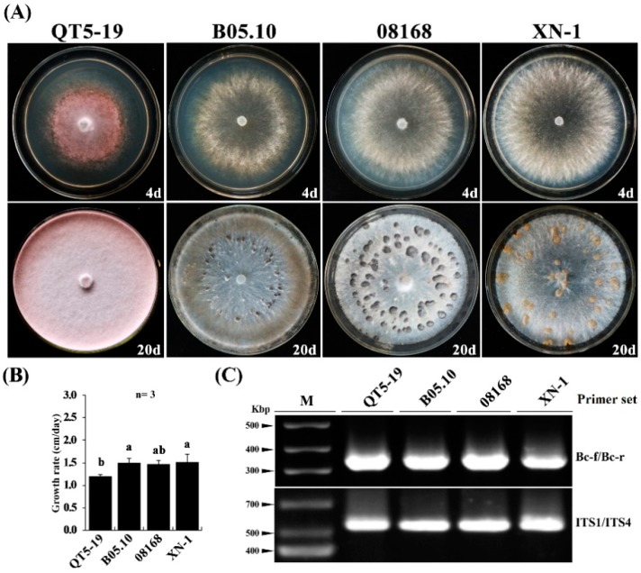
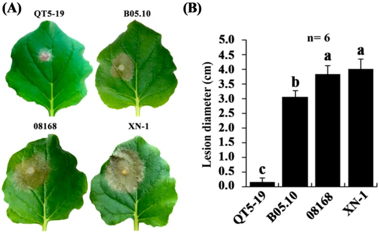
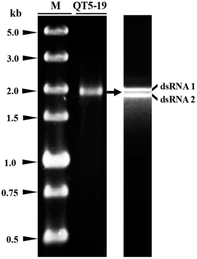
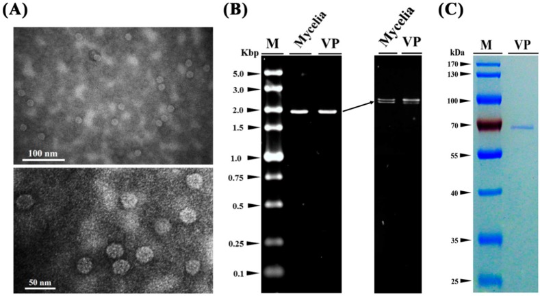
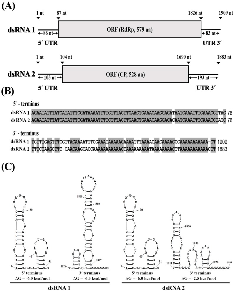
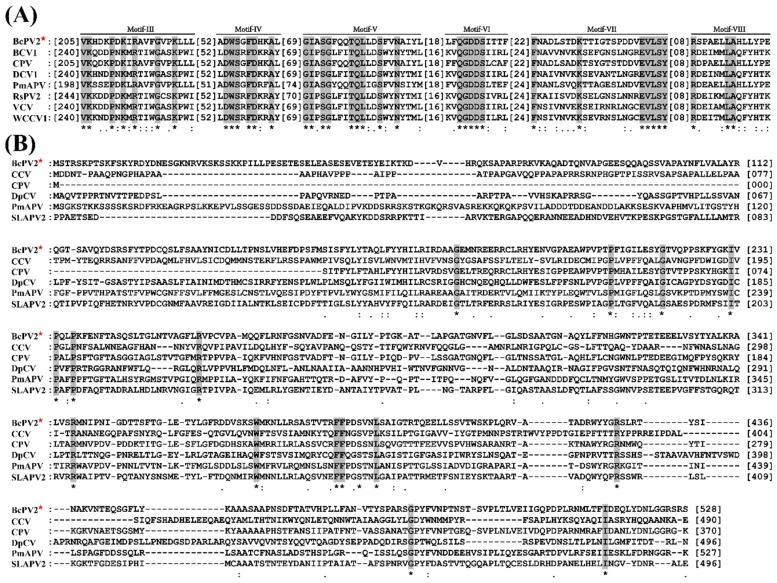
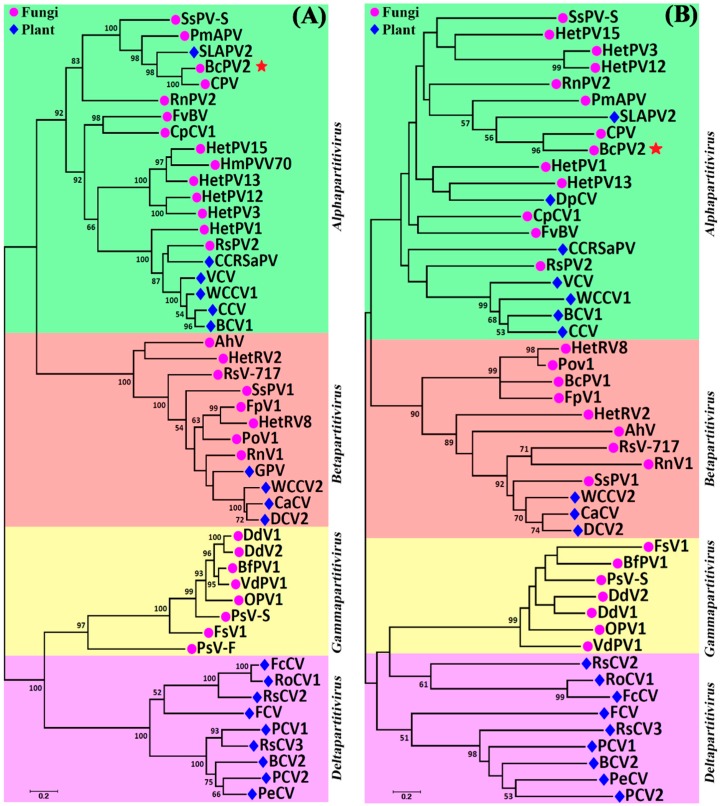
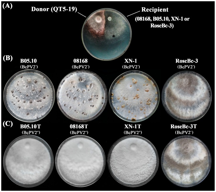
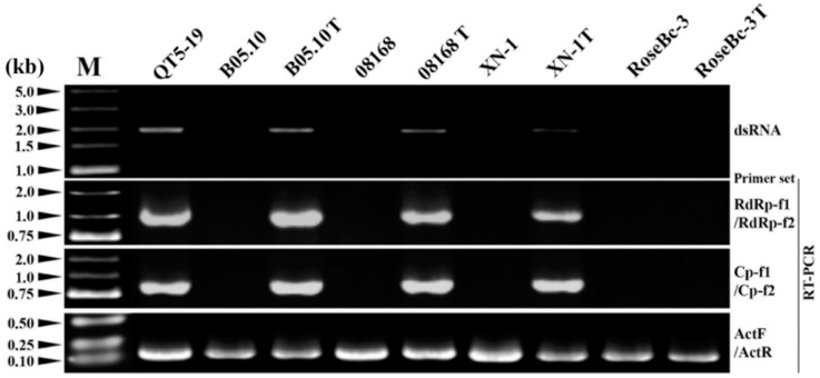
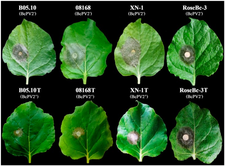
A pink isolate (QT5-19) of Botrytis cinerea was compared with three gray isolates of B. cinerea for growth and morphogenesis on potato dextrose agar (PDA), and for pathogenicity on tobacco. A double-stranded (ds) RNA mycovirus infecting QT5-19 was identified based on its genome feature and morphology of the virus particles. The results showed that QT5-19 grew rapidly and established flourishing colonies as the gray isolates did. However, it is different from the gray isolates, as it failed to produce conidia and sclerotia asthe gray isolates did. QT5-19 hardly infected tobacco, whereas the gray isolates aggressively infected tobacco. Two dsRNAs were detected in QT5-19, dsRNA 1 and dsRNA 2, were deduced to encode two polypepetides with homology to viral RNA-dependent RNA polymerase (RdRp) and coat protein (CP), respectively. Phylogenetic analysis of the amino acid sequences of RdRp and CP indicated that the two dsRNAs represent the genome of a novel partitivirus in the genus Alphapartitivirus, designated here as Botrytis cinerea partitivirus 2 (BcPV2). BcPV2 in QT5-19 was successfully transmitted to the three gray isolates through hyphal contact. The resulting BcPV2-infected derivatives showed rapid growth on PDA with defects in conidiogenesis and sclerogenesis, and hypovirulence on tobacco. This study suggests that BcPV2 is closely associated with hypovirulence of B. cinerea.

Keywords: Botrytis cinerea; conidiogenesis; hypovirulence; partitivirus; sclerogenesis.


Conflict of interest statement

The authors declare no conflict of interest.

Figures

Figure 1
Figure 1
Cultural characteristics of Botrytis cinerea isolates on potato dextrose agar. (A) Four-day-old (top) and 20-day-old cultures (bottom) of isolates QT5-19, 08168, B05.10, and XN-1 (20 °C). Note difference in colony color, pink for QT5-19, whereas gray for the other three isolates. Note also difference in sclerotial color, orange sclerotia produced by isolate XN-1, whereas black sclerotia produced by isolates 08168 and B05.10; (B) A histogram showing average mycelial growth rates of the four isolates. Means ± S.D. (n = 4) labeled with the same letters are not significantly different at p > 0.05; (C) PCR-based identification of QT5-19 using the B. cinerea-specific primer set Bc-f/Bc-r and the universal primer set ITS1/ITS4. Isolates 08168, B05.10, and XN-1 of B. cinerea were used as reference.
Figure 2
Figure 2
Pathogenicity of Botrytis cinerea isolates on tobacco (Nicotiana benthamiana). (A) Four tobacco leaves inoculated with the mycelia of the isolates QT5-19, 08168, B05.10, and XN-1, respectively (20 °C, 3 dpi). Note no visible lesion formation on the leaf inoculated with isolate QT5-19, whereas formation of large necrotic lesions on the leaves inoculated with the other three isolates; (B) A histogram showing average leaf lesion diameters caused by the four isolates. Means ± S.D. (n = 6) labeled with the same letters are not significantly different at p > 0.05.
Figure 3
Figure 3
Double-stranded (ds) RNAs in isolate QT5-19 of Botrytis cinerea. The electrophoregram on the left was carried out in a 1.0% agarose gel after 1.5-h electrophoresis under room temperature. The electrophoregram on the right was carried out in a 0.7% agarose gel after 15-h electrophoresis at 4 °C. The dsRNAs were treated with RNase-free DNase I and S1 nuclease before electrophoresis. M = DL5000 dsDNA marker (TaKaRa).
Figure 4
Figure 4
Virus particles isolated from the mycelia of isolate QT5-19 of Botrytis cinerea. (A) Two transmission electron microscope (TEM) graphs showing the shape and size of the virus particles of the mycovirus in QT5-19; (B) Electrophoregrams showing the two dsRNAs extracted from the virus particles (VP) and the mycelia of QT5-19 (Mycelia). The electrophoregram on the right was created in a 1% agarose gel after 1.5-h electrophoresis under room temperature. The electrophoregram on the left was created in a 0.7% agarose gel after 15-h electrophoresis at 4 °C. M = DL5000 marker (TaKaRa); (C) An SDS-PAGE (10%) electrophoregram showing the band of the structural protein (coat protein) extracted from the virus particles of the mycovirus in QT5-19. The gel was stained with Coomassie brilliant blue R-250. M = PageRuler™ Prestained Protein Ladder.
Figure 5
Figure 5
Genome structure of BcPV2. (A) A schematic diagram illustrating the segmented dsRNA genome. Each dsRNA has one open reading frame (ORF) flanked by two untranslated regions (UTR) at 3′- and 5′-termini; (B) Alignments of the partial nucleotide sequences of the 3′- and 5′-UTRs of dsRNA 1 and dsRNA 2. The identical nucleotides were highlighted in gray color. “-”, missing nucleotides; (C) Predicted secondary structures for the terminal regions of dsRNA 1 and dsRNA 2.
Figure 6
Figure 6
Multiple alignments of the amino acid sequences of RNA-dependent RNA polymerase and coat protein of BcPV2 with other selected members in Alphapartitivirus. (A) Amino acid sequences of RNA-dependent RNA polymerase (RdRp); and (B) amino acid sequences of coat protein (CP). “*”, identical amino acids, “:” and “.” high and low chemically similar amino acids, respectively. Note the five conserved motifs (Motif-III to Motif-VIII) in RdRp and diversified CP sequences among the compared viruses. Abbreviations: BCV1, Beet cryptic virus 1; BcPV2, Botrytis cinerea partitivirus 2; CCV, carrot cryptic virus; CPV, Ceratobasidium partitivirus; DCV1, Dill cryptic virus 1; DpCV, Diuris pendunculata cryptic virus; PmAPV, Powdery mildew-associated partitivirus; RsPV2, Rhizoctonia solani partitivirus 2; SLAPV2, Soybean leaf-associated partitivirus 2; VCV, Vicia cryptic virus; WCCV1, white clover cryptic virus 1. The GenBank accession numbers for RdRp and CP of the above-mentioned viruses are listed in Table S2.
Figure 7
Figure 7
Two phylogenetic trees showing the relationship of BcPV2 with other partitiviruses. The trees were inferred based on amino acid sequences of RdRp (A) and CP (B) using the Maximum-Likelihood method with bootstrap values determined by 1000 replicates. Bootstrap values higher than 50% are shown in the graphs. The scale bars represent substitutions per nucleotide position. Abbreviations: AhV, Atkinsonella hypoxylon partitivirus; BcPV1, Botrytis cinerea Partitivirus 1; BcPV2, Botrytis cinerea Partitivirus 2; BCV1, Beet cryptic virus 1; BCV2, Beet cryptic virus 2; BfPV1, Botryotinia fuckeliana partitivirus 1; CaCV, Cannabis cryptic virus; CCRSaPV, Cherry chlorotic rusty spot associated partitivirus; CpCV1, Chondrostereum purpureum cryptic virus 1; CPV, Ceratobasidium partitivirus; DCV1, Dill clover cryptic virus 1; DCV2, Dill cryptic virus 2; DdV1, Discula destructiva virus 1; DdV2, Discula destructiva virus 2; FcCV, Fragaria chiloensis cryptic virus; FCV, Fig cryptic virus; FpV1, Fusarium poae virus 1; FsV1, Fusarium solani virus 1; FvBV, Flammulina velutipes browning virus; GPV, Grapevine partitivirus; HetPV1, Heterobasidion partitivirus 1; HetPV12, Heterobasidion partitivirus 12; HetPV13, Heterobasidion partitivirus 13; HetPV15, Heterobasidion partitivirus 15; HetPV3, Heterobasidion partitivirus 3; HetRV2, Heterobasidion partitivirus 2; HetRV8, Heterobasidion partitivirus 8; HmPVV70, Helicobasidium mompa partitivirus V70; OPV1, Ophiostoma partitivirus 1; PCV1, Pepper cryptic virus 1; PCV2, Pepper cryptic virus 2; PeCV, Persimmon cryptic virus; PmAPV, Powdery mildew-associated partitivirus; PoV1, Pleurotus ostreatus virus 1; PsV-F, Penicillium stoloniferum virus F; PsV-S, Penicillium stoloniferum virus S; RnPV2, Rosellinia necatrix partitivirus 2; RnV1, Rosellinia necatrix partitivirus 1; RoCV1, Rose cryptic virus 1; RsCV2, Raphanus sativus cryptic virus 2; RsCV3, Raphanus sativus cryptic virus 3; RsPV2, Rhizoctonia solani partitivirus 2; RsV-717, Rhizoctonia solani virus 717; SLAPV2, Soybean leaf-associated partitivirus 2; SsPV1, Sclerotinia sclerotiorum partitivirus 1; SsPV-S, Sclerotinia sclerotiorum partitivirus S; VCV, Vicia cryptic virus; VdPV1, Verticillium dahliae partitivirus 1; WCCV1, White clover cryptic virus 1; WCCV2, White clover cryptic virus 2. The GenBank accession number of the above-mentioned viruses are listed in Table S2.
Figure 8
Figure 8
Horizontal transmission of BcPV2 through hyphal contact in pair cultures on PDA. (A) A pair culture on PDA with the colonies of isolate QT5-19 (BcPV2 donor, pink color) and a recipient (gray color). The symbol (*) in the colony indicated the area where a mycelial agar plug was removed and transferred to PDA for establishing a BcPV2-transmitted derivative; (B) Four PDA cultures (20 °C, 20 day) of the B. cinerea isolates free of infection by BcPV2 (BcPV2); (C) Four PDA cultures (20 °C, 20 day) of the transfected derivatives of B. cinerea. BcPV2+, infected by BcPV2, BcPV2, not infected by BcPV2.
Figure 9
Figure 9
Detection of BcPV2 in different isolates of Botrytis cinerea by dsRNA profiling and RT-PCR with specific primers. M = DL5000 DNA marker (TaKaRa). Isolates B05.10T, 08168T, XN-1T, and RoseBc-3T were derived from B05.10, 08168, XN-1 and RoseBc-3 in the pairing cultures of QT5-19/B05.10, QT5-19/08168, QT5-19/XN-1 and QT5-19/RoseBc-3, respectively.
Figure 10
Figure 10
Pathogenicity of different isolates of Botrytis cinerea on detached leaves of Nicotiana benthamiana (20 °C, 72 h). Note smaller leaf lesion size caused by B05.10T, 08168T, and XN-1T than that caused by B05.10, 08168 and XN-1, whereas similar leaf lesion size caused by RoseBc-3T and RoseBc-3. BcPV+ = infected by BcPV2; BcPV2 = not infected by BcPV2.

References

    1. Ghabrial S.A., Suzuki N. Viruses of plant pathogenic fungi. Annu. Rev. Phytopathol. 2009;47:353–384. doi: 10.1146/annurev-phyto-080508-081932. - DOI - PubMed
    1. Pearson M.N., Beever R.E., Boine B., Arthur K. Mycoviruses of filamentous fungi and their relevance to plant pathology. Mol. Plant Pathol. 2009;10:115–128. doi: 10.1111/j.1364-3703.2008.00503.x. - DOI - PMC - PubMed
    1. Herrero N., Márquez S.S., Zabalgogeazcoa I. Mycoviruses are common among different species of endophytic fungi of grasses. Arch. Virol. 2009;154:327–330. doi: 10.1007/s00705-008-0293-5. - DOI - PubMed
    1. Ghabrial S.A., Castón J.R., Jiang D., Nibert M.L., Suzuki N. 50-plus years of fungal viruses. Virology. 2015;479:356–368. doi: 10.1016/j.virol.2015.02.034. - DOI - PubMed
    1. Xie J., Jiang D. New insights into mycoviruses and exploration for the biological control of crop fungal diseases. Annu. Rev. Phytopathol. 2014;52:45–68. doi: 10.1146/annurev-phyto-102313-050222. - DOI - PubMed

Publication types