Review
. 2019 Jan 3;12(1):136.
doi: 10.3390/ma12010136.

Magnesium Implants: Prospects and Challenges

Affiliations
Review

Magnesium Implants: Prospects and Challenges

Parama Chakraborty Banerjee et al. Materials (Basel). .

Abstract

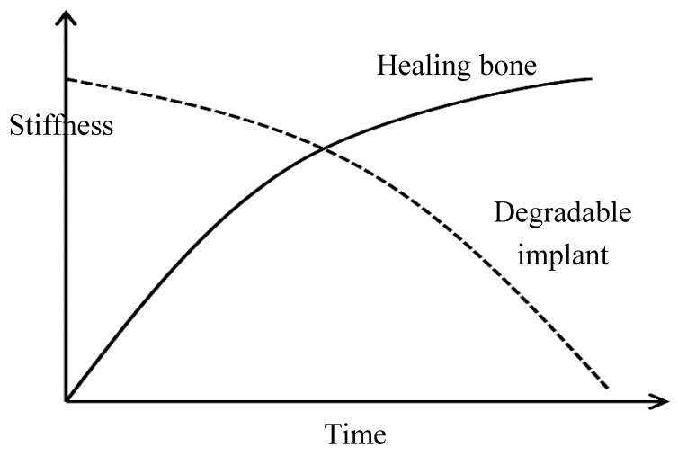
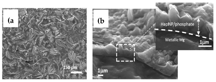
Owing to their suitable mechanical property and biocompatibility as well as the technological possibility of controlling their high corrosion rates, magnesium and its alloys have attracted significant attention as temporary bio-implants. Though the ability of magnesium to harmlessly biodegrade and its inherent biocompatibility make magnesium alloys a suitable choice for a temporary implant, their high corrosion rates limit their practical application, as the implants can potentially corrode away even before the healing process has completed. Different approaches, such as alloying, surface modification, and conversion coatings, have been explored to improve the corrosion resistance of various magnesium alloys. However, the corrosion behavior of magnesium implants with and without a surface modification has been generally investigated under in-vitro conditions, and studies under in-vivo conditions are limited, which has contributed to the lack of translation of magnesium implants in practical applications. This paper comprehensively reviews the prospects of magnesium alloy implants and the current challenges due to their rapid degradation in a physiological environment. This paper also provides a comprehensive review of the corrosion mitigation measures for these temporary implants.

Keywords: corrosion; implant; magnesium alloy.


Conflict of interest statement

The authors declare no conflicts of interest.

Figures

Figure 1
Figure 1
The gradual decrease in stiffness of a biodegradable magnesium implant and concurrent healing of the bone. Obtained from [13], with permission from Woodhead Publishing, 2018.
Figure 2
Figure 2
Radiographs of hip implants showing changes in bone mass due to stress shielding around an implant: (a) bone loss at the proximal region (indicated in the circled regions at the edge of the prosthesis) and (b) bone deposition (indicated by the rectangle) in the distal region of the implant . Obtained from [23], with permission from Wiley online library, 2018.
Figure 3
Figure 3
The increase in corrosion resistance of the poly-lactic acid (PLA)-coated AZ21 alloy with increasing coating thickness. Obtained from [65], with permission from Thin Solid Films, Elsevier, 2018.
Figure 4
Figure 4
The magnesium ion concentration of the poly (lactic-co-glycolic acid) polymer-coated (a) AZ21 and (b) Mg4Y alloy substrates in DMEM at various days of immersion. Obtained from [65], with permission from Thin Solid Films, Elsevier, 2018.
Figure 4
Figure 4
The magnesium ion concentration of the poly (lactic-co-glycolic acid) polymer-coated (a) AZ21 and (b) Mg4Y alloy substrates in DMEM at various days of immersion. Obtained from [65], with permission from Thin Solid Films, Elsevier, 2018.
Figure 5
Figure 5
Surface morphology (a,c,e) and cross-section images (b,d,f) of the (a,b) micro-arc oxidation (MAO)-treated, (c,d) MAO-4% PCL duplex-coated, and (e,f) MAO-7% PCL duplex-coated magnesium. Obtained from [67], with permission from Thin Solid Films, Elsevier, 2018.
Figure 6
Figure 6
The potentiodynamic polarization of the uncoated, hydroxide pre-treated, BTSE-coated, γ-APS-coated, BTSE/γ-APS-coated, and BTSE/γ-APS/heparin-coated AZ31 alloy in simulated body fluid. Obtained from [78], with permission from Acta Biomaterialia, Elsevier, 2018.
Figure 7
Figure 7
Surface morphologies of: (a) brushite-coated, (b) hydroxyapatite-coated, and (c) fluoridated-hydroxyapatite-coated Mg–Zn alloy substrates. (d) Cross-sectional view of the fluoridated-hydroxyapatite-coated specimen. Obtained from [84], with permission from Acta Biomaterialia, Elsevier, 2018.
Figure 8
Figure 8
Surface morphologies of the Ca–P coating synthesized using (a,c) constant potential and (b,d) pulse potential techniques. Obtained from [87], with permission from Materials Letters, Elsevier, 2018.
Figure 9
Figure 9
Post-corrosion morphologies of the (a,b) uncoated and (c,d) nanostructured-hydroxyapatite-coated alloy substrate after 7 days of immersion in simulated body fluid. Obtained from [91], with permission from Materials Science and engineering C, Elsevier, 2018.
Figure 10
Figure 10
(a) open circuit voltage versus time and (b) potentiodynamic polarization plots of the uncoated, hydroxyapatite-coated, and graphene oxide (GO)/hydroxyapatite-coated titanium substrate. Obtained from [101], with permission from Carbon, Elsevier, 2018.
Figure 11
Figure 11
Surface morphologies of (a) the graphene oxide coating on AZ91. (bd) the graphene oxide/hydroxyapatite coating with a growth time of 6 h, 1 day, and 2 days, respectively, at a pH of 6.65, (eg) the hydroxyapatite-only coating with a growth time of 6 h, 1 day, and 2 days, respectively, at a pH of 6.65. A graphene oxide/hydroxyapatite coating grown for 2 days at a pH of (h) 6.4 and (i) 6.9. Obtained from [102], with permission from Materials Letters, Elsevier, 2018.
Figure 12
Figure 12
The (a) surface morphology and (b) cross section of a graphene oxide/hydroxyapatite nanoparticle coating on a high-purity magnesium substrate. Obtained from [103], with permission from Applied Surface Science, Elsevier, 2018.

References

    1. Mushahary D. Surface Functionalisation of Magnesium Alloys for Use as Bio-Implants. Institute for Frontier Materials, Deakin University; Geelong, Australia: 2014.
    1. Niinomi M. Recent metallic materials for biomedical applications. Metall. Mater. Trans. A. 2002;33:477. doi: 10.1007/s11661-002-0109-2. - DOI
    1. Williams D.F. Corrosion of Implant Materials. Annu. Rev. Mater. Sci. 1976;6:237–266. doi: 10.1146/annurev.ms.06.080176.001321. - DOI
    1. Nielsen K. Corrosion of metallic implants. Br. Corros. J. 1987;22:272–278. doi: 10.1179/000705987798271352. - DOI
    1. Niinomi M. Metallic biomaterials. J. Artif. Organs. 2008;11:105. doi: 10.1007/s10047-008-0422-7. - DOI - PubMed

LinkOut - more resources