. 2019 Jan 3;11(1):39.
doi: 10.3390/cancers11010039.

Prognostic Impact of Tumor-Infiltrating Lymphocytes and Neutrophils on Survival of Patients with Upfront Resection of Pancreatic Cancer

Affiliations

Prognostic Impact of Tumor-Infiltrating Lymphocytes and Neutrophils on Survival of Patients with Upfront Resection of Pancreatic Cancer

Rainer C Miksch et al. Cancers (Basel). .

Abstract

In patients with pancreatic ductal adenocarcinoma (PDAC), the tumor microenvironment consists of cellular and stromal components that influence prognosis. Hence, tumor-infiltrating lymphocytes (TILs) may predict prognosis more precisely than conventional staging systems. Studies on the impact of TILs are heterogeneous and further research is needed. Therefore, this study aims to point out the importance of peritumoral TILs, tumor-infiltrating neutrophils (TINs), and immune subtype classification in PDAC. Material from 57 patients was analyzed with immunohistochemistry performed for CD3, CD8, CD20, CD66b, α-sma, and collagen. Hot spots with peritumoral TILs and TINs were quantified according to the QTiS algorithm and the distance of TILs hot spots to the tumor front was measured. Results were correlated with overall (OS) and progression-free survival (PFS). High densities of peritumoral hot spots with CD3⁺, CD8⁺, and CD20⁺ TILs correlated significantly with improved OS and PFS. Combined immune cell subtypes predicted improved OS and PFS. High infiltration of CD3⁺ TILs predicted progression after 12 months. The location of TILs' hot spots and their distance to the tumor front did not correlate with patient survival. Peritumoral TILs and the composition of the stroma predict OS and PFS in PDAC.

Keywords: activated stroma index; immune cell infiltration; immune infiltrate; immunoscore; immunosuppression; pancreatic ductal adenocarcinoma; quantification of the tumor immune stroma (QTiS); tumor microenvironment; tumor-infiltrating lymphocytes.


Conflict of interest statement

The authors declare no conflict of interest.

Figures
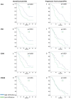
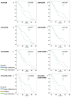
Figure 1
Figure 1
Survival graphs of overall (AD) and progression-free survival (EH) for high (blue) vs. low (green) infiltration of CD3+, CD8+, CD20+, and CD66b+ tumor-infiltrating leukocytes (TILs). The median value was used to distinguish high vs. low infiltration after quantification of stained cells [39]. High infiltration with CD3+, CD8+, and CD20+ TILs correlated significantly with improved OS and PFS. No statistically significant influence was evident for CD66b+ TINs.
Figure 2
Figure 2
Survival graphs of OS in relation to different combinations of TILs and TINs: Groups were formed by separating high infiltration in both cell types (blue color, e.g., high CD3+ and high CD8+) from the rest (green color, e.g., high/low CD3+/CD8+ or low CD3+ and low CD8+), where two immune cells were analyzed together (AF). Groups were formed by separating high infiltration in all cell types, low infiltration in all cell types, and mixed levels of infiltration, where three immune cells were analyzed together (G). Finally, combination with four immune cells was performed (H). High infiltration of CD3+/CD8+, CD3+/CD8+/CD20+, CD8+/CD20+, CD20+/CD66b+, and CD3+/CD8+/CD20+/CD20+/CD66b+ tumor-infiltrating leukocytes correlated significantly with better OS.
Figure 3
Figure 3
Survival graphs of PFS in relation to different combinations of TILs and TINs: Groups were formed by separating high infiltration in both cell types accordingly to Figure 2. (AF) Groups were combined with two cell types, (G) with three, and (H) with four different cell types. High infiltration of CD3+/CD8+, CD3+/CD8+/CD20+, CD8+/CD20+, CD20+/CD66b+, and CD3+/CD8+/CD20+/CD20+/CD66b+ tumor-infiltrating leukocytes correlated significantly with better PFS.
Figure 4
Figure 4
Kaplan-Meier survival curves of overall (A,C,E) and progression-free survival (B,D,F) in relation to the distance in µm of CD3+ (A,B), CD8+ (C,D) or CD20+ (E,F) hot spots from the tumor front. The groups are divided into hot spots close to the tumor front (blue) and hot spots far from tumor front (green) according to the median distance value: 0.0333253 µm for CD3, 0.0687577 µm for CD8, and 0.056765 µm for CD20.
Figure 5
Figure 5
Hot spots with tumor-infiltrating leukocytes of CD3+ (A), CD8+ (B), CD20+ (C), and CD66b+ (D) cells under a power field of 200× magnification as well as corresponding areas of α-sma (E) and collagen (F) stained spots. CD3 is the marker of T cells, CD8—of cytotoxic T cells, CD20—of B cells, CD66b—of neutrophils, and α-sma—of cancer associated fibroblasts.

Similar articles

Cited by

References

    1. Siegel R., Naishadham D., Jemal A. Cancer statistics, 2012. CA Cancer J. Clin. 2012;62:10–29. doi: 10.3322/caac.20138. - DOI - PubMed
    1. Qiu D., Kurosawa M., Lin Y., Inaba Y., Matsuba T., Kikuchi S., Yagyu K., Motohashi Y., Tamakoshi A. Overview of the epidemiology of pancreatic cancer focusing on the JACC Study. J. Epidemiol. 2005;15(Suppl. 2):S157–S167. doi: 10.2188/jea.15.S157. - DOI - PMC - PubMed
    1. Talamini G., Bassi C., Falconi M., Sartori N., Salvia R., Rigo L., Castagnini A., Di Francesco V., Frulloni L., Bovo P., et al. Alcohol and smoking as risk factors in chronic pancreatitis and pancreatic cancer. Dig. Dis. Sci. 1999;44:1303–1311. doi: 10.1023/A:1026670911955. - DOI - PubMed
    1. Yadav D., Whitcomb D.C. The role of alcohol and smoking in pancreatitis. Nat. Rev. Gastroenterol. Hepatol. 2010;7:131–145. doi: 10.1038/nrgastro.2010.6. - DOI - PubMed
    1. Raimondi S., Maisonneuve P., Lowenfels A.B. Epidemiology of pancreatic cancer: An overview. Nat. Rev. Gastroenterol. Hepatol. 2009;6:699–708. doi: 10.1038/nrgastro.2009.177. - DOI - PubMed