. 2019 Jan 4;18(1):1.
doi: 10.1186/s12943-018-0930-x.

Epigenetically upregulated oncoprotein PLCE1 drives esophageal carcinoma angiogenesis and proliferation via activating the PI-PLCε-NF-κB signaling pathway and VEGF-C/ Bcl-2 expression

Affiliations

Epigenetically upregulated oncoprotein PLCE1 drives esophageal carcinoma angiogenesis and proliferation via activating the PI-PLCε-NF-κB signaling pathway and VEGF-C/ Bcl-2 expression

Yunzhao Chen et al. Mol Cancer. .

Abstract

Background: Esophageal squamous cell carcinoma (ESCC) is one of the most lethal malignancies. Neovascularization during tumorigenesis supplies oxygen and nutrients to proliferative tumor cells, and serves as a conduit for migration. Targeting oncogenes involved in angiogenesis is needed to treat organ-confined and locally advanced ESCC. Although the phospholipase C epsilon-1 (PLCE1) gene was originally identified as a susceptibility gene for ESCC, how PLCE1 is involved in ESCC is unclear.

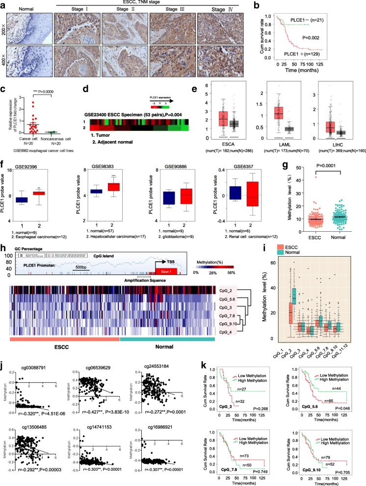
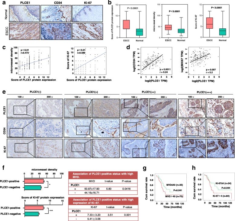
Methods: Matrix-assisted laser desorption ionization time-of-flight mass spectrometry were used to measure the methylation status of the PLCE1 promoter region. To validate the underlying mechanism for PLCE1 in constitutive activation of the NF-κB signaling pathway, we performed studies using in vitro and in vivo assays and samples from 368 formalin-fixed esophageal cancer tissues and 215 normal tissues with IHC using tissue microarrays and the Cancer Genome Atlas dataset.

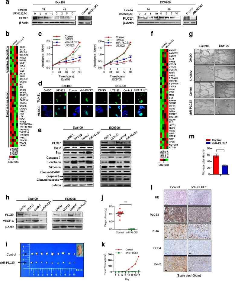
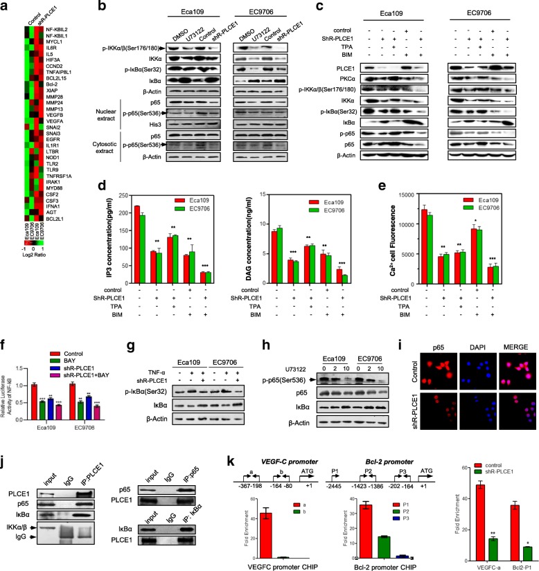
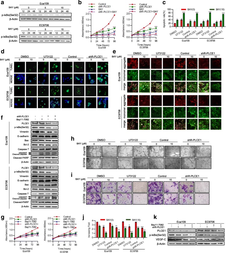
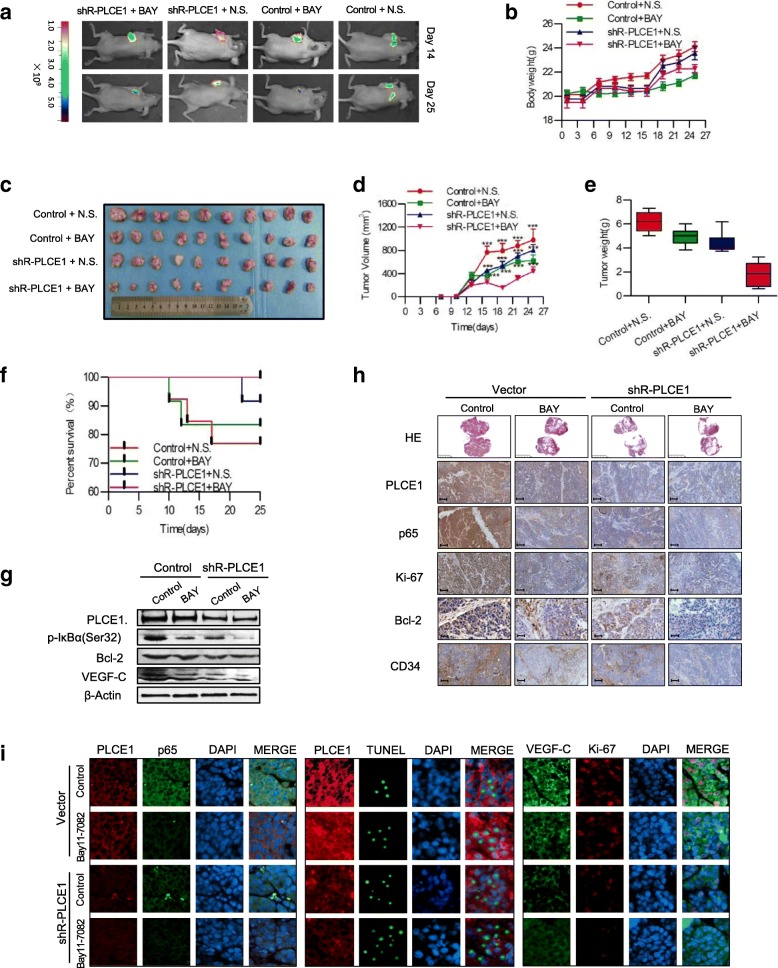
Results: We report that hypomethylation-associated up-regulation of PLCE1 expression was correlated with tumor angiogenesis and poor prognosis in ESCC cohorts. PLCE1 can activate NF-κB through phosphoinositide-phospholipase C-ε (PI-PLCε) signaling pathway. Furthermore, PLCE1 can bind p65 and IκBα proteins, promoting IκBα-S32 and p65-S536 phosphorylation. Consequently, phosphorylated IκBα promotes nuclear translocation of p50/p65 and p65, as a transcription factor, can bind vascular endothelial growth factor-C and bcl-2 promoters, enhancing angiogenesis and inhibiting apoptosis in vitro. Moreover, xenograft tumors in nude mice proved that PLCE1 can induce angiogenesis, inhibit apoptosis, and increase tumor aggressiveness via the NF-κB signaling pathway in vivo.

Conclusions: Our findings not only provide evidence that hypomethylation-induced PLCE1 confers angiogenesis and proliferation in ESCC by activating PI-PLCε-NF-κB signaling pathway and VEGF-C/Bcl-2 expression, but also suggest that modulation of PLCE1 by epigenetic modification or a selective inhibitor may be a promising therapeutic approach for the treatment of ESCC.

Keywords: Angiogenesis; Esophageal carcinoma; NF-κB; PLCE1; Proliferation.


Conflict of interest statement

Ethics approval and consent to participate

All experiments were approved by the Ethics Committee of The First Affiliated Hospital, Shihezi University School of Medicine.

Consent for publication

All authors give consent for the publication of the manuscript in Molecular Cancer.

Competing interests

The authors declare that they have no competing interests.

Publisher’s Note

Springer Nature remains neutral with regard to jurisdictional claims in published maps and institutional affiliations.

Figures

Fig. 1
Fig. 1
PLCE1 is upregulated in ESCC through aberrant promoter hypomethylation. a IHC staining of PLCE1 expression in human ESCC (clinical stages I–IV) and normal esophageal tissues. b Kaplan-Meier overall survival curves for patients with ESCC stratified by low (n = 21) and high (n = 129) expressions of PLCE1 (P = 0.002). c GEO data (GSE9982) analysis of PLCE1 mRNA expression in normal esophageal cell lines (n = 20) and esophageal cancer cell lines (n = 20). ***P < 0.0009, unpaired two-tailed Student’s t-test. d Analysis of PLCE1 gene expression in tumor and adjacent non-tumor tissues in NCBI/GEO/GSE23400 (P = 0.004). e GEPIA analysis of PLCE1 expression in cancerous and normal tissues. ESCA: esophageal carcinoma; LAML: Acute Myeloid Leukemia; LIHC: Liver hepatocellular carcinoma; *P < 0.05. f GEO data analysis for the expression of PLCE1 by two-tailed t test (*P < 0.05, **P < 0.01, ***P < 0.001). g Genomic structure of PLCE1 CpG dinucleotides over TSS and hierarchical cluster analysis of CpG units’ methylation profiles of PLCE1 promoter region in ESCC (n = 132) and normal (n = 104). Each vertex indicates one CpG site. Each column represents one sample. Rows are clustering of CpG units, which are single CpG sites or a combination of CpG sites. Color gradient between white and red indicates methylation of each PLCE1 CpG unit in each sample (0–56%). Black represents inadequate or missing data. h Comparison of average methylation of PLCE1 promoter of ESCC and normal subjects. The overall methylation level of the target fragment of the PLCE1 promoter was statistically lower (0.0957 ± 0.0456) in ESCC than that in normal tissues (0.1144 ± 0.0464, P = 0.0001). i Box plot of 6 CpG units in PLCE1 promoter between ESCC and normal tissues. Red and blue spots represent methylation status of one CpG site in ESCC and normal tissues. Dark spots are outliers. In addition to CpG_3 an d CpG_4, the mean methylation levels at CpG_2, CpG_5.6, CpG_7.8, and CpG_9.10 were significantly lower in patients with ESCC (mean methylation = 20.25, 11.84, 8.07, 7.20%, respectively) than those in the controls (mean methylation = 32.38, 13.89, 10.52, 9.37%, respectively; all the P < 0.05), *P < 0.05, **P < 0.01, ***P < 0.001 (Mann–Whitney U-test). j The methylation status of 6 CpG site was all negatively correlated with PLCE1 expression in TCGA Illumina 450 k infinium methylation beadchip. k Kaplan-Meier analysis of survival time according to PLCE1 CpG methylation in ESCC patients
Fig. 2
Fig. 2
Overexpression of PLCE1 is relevant with the angiogenesis and proliferation of ESCC. a Representative IHC images of PLCE1, CD34 and Ki-67 staining (200×) in ESCC and normal esophageal tissue specimens. b The score of PLCE1 and Ki-67 and the MVD in ESCC were higher than normal esophageal tissues. Each bar represents the mean ± SD for triplicate experiments. c, d The correlation analysis between PLCE1 with MVD and Ki-67 were carried out. PLCE1 and MVD are positively correlated, so as PLCE1 and Ki-67. e Representative IHC images of PLCE1, CD34 and Ki-67 staining according to intensities of PLCE1, CD34 and Ki-67 staining in consecutive ESCC tissues. f MVD numbers and Ki-67 score were higher in IHC with positive PLCE1 expression compared to negative c-kit expression. g, h Kaplan-Meier curves for patients. High-MVD (MVD > 40) group (n = 70) and Ki-67-positive (n = 84) had poor prognosis compared with low-MVD expression group (n = 35) and a Ki-67-negative group (n = 34)
Fig. 3
Fig. 3
PLCE1 promotes proliferation and angiogenesis of ESCC cells in vitro and induces aggressiveness in vivo. a Eca109 and EC9706 cells treated with U73122 at 0, 2, 5, and 10 concentrations for 24 and 48 h. ShR-PLCE1 were transfected at a MOI of 15 for 60 h. PLCE1 expression measured by Western blot. b Real-time PCR analysis demonstrating relationship between PLCE1 expression and apoptosis. Color represents intensity scale for vector of PLCE1 shRNA versus control, as calculated by log2 transformation. c Eca109 and EC9706 cells treated shR-PLCE1 or U73122 at the indicated concentration for 0, 24, 48, 72, and 96 h. Cell viability measured by MTT and presented as means ± SD from three separate experiments. d TUNEL staining of cells treated as indicated; e shR-PLCE1 on apoptosis-related proteins assayed by Western blot. β-Actin was a loading control. f Real-time PCR analysis demonstrated the positive relationship between PLCE1 expression and angiogenesis. Pseudo-color represents intensity scale for the vector or PLCE1 shRNA versus control, as calculated by log2 transformation. g Tube formation by indicated cells. h Effects of shR-PLCE1 on VEGF-C protein expression as detected by Western blot. i Xenograft model in nude mice; representative images of tumors from all mice in each group. Mean tumor weights j and tumor volume growth curves k for tumors formed by the indicated cells. l H&E and IHC staining demonstrated that PLCE1 induced the aggressive phenotype of ESCC cells in vivo. Scale bar, 100 μm. Microvascular density m show that PLCE1 promotes resistance to apoptosis and angiogenesis in vivo. All data are presented as mean ± SD. *P < 0.05, ***P < 0.001
Fig. 4
Fig. 4
PLCE1 activates the NF-κB signaling pathway in ESCC. a Real-time PCR analysis shows overlap between NF-κB-dependent gene expression and PLCE1-regulated gene expression. Color represents intensity for vector of PLCE1 shRNA versus control, as calculated by log2 transformation. b Western blot for indicated proteins in indicated cells. c Western blot analysis was performed to indicate PLCE1 affect NF-κB signaling pathway through PI-PLCε pathway by using PLCE1 shRNA, TPA, and BIM. d ELISA assays showed the effects of PLCE1 shRNA, TPA, and BIM on intracellular levels of the PI-PLCε-pathway-related proteins IP3 and DAG. e Laser scanning confocal microscopy was used to measure intracellular calcium fluorescence pixel values of ESCC cells after treatment with PLCE1 shRNA, TPA, and BIM, which triggered positive and negative effects on the PI-PLC pathway. The histogram shows semi-quantitative analysis of fluorescence. f Activity of NF-κB luciferase reporter gene in ESCC cells expressed with indicated treatment. g IκBα and p-IκBα (Ser 32) in indicated cells. h Effects of 48 h U73122 treatment at 0, 2, and 10 concentrations on protein levels of phosphorylated p65 (Ser536); p65 and IκBα in ESCC cells. i IF analysis of Eca109 and EC9706 cells transfected with shR-PLCE1. Cells were fixed, stained with antibodies to p65 (red) and by 4′,6-diamidino-2-phenylindole (blue), incubated with appropriate secondary antibodies, and analyzed using double IF assays. j Whole cell lysates from Eca109 and EC9706 cells immunoprecipitated with antibodies against indicated proteins. k ChIP assay p65-binding sites on Bcl2 and VEGF-C genes. Extent of recruitment assessed by real-time PCR
Fig. 5
Fig. 5
PLCE1 inhibits apoptosis and enhances angiogenesis via activation of the NF-κB signaling pathway in ESCC in vitro. a Eca109 and EC9706 cells treated with Bay11–7082 at the 0, 5, 15 concentrations for 12, 24 and 48 h. p-IκBα(Ser32) expression measured by Western blot. b Eca109 and EC9706 cells were treated with shR-PLCE1 or/and Bay11–7082 at the indicated concentration for 0, 24, 48, 72, and 96 h. Cell viability was measured by MTT and presented as means ± SD from three separate experiments. c FITC–PI staining of cells treated with shR-PLCE1 or U73122 or/and Bay11–7082 and results means ± SD from three independent experiments. d TUNEL staining of cells treated with indicated concentration. e Eca109 and EC9706 cells were treated with shR-PLCE1 or U73122 or/and Bay11–7082 and then subjected to JC-1 staining assays. f Effects of shR-PLCE1 and Bay11–7082 on apoptosis-related proteins assayed by Western blot. β-Actin was loading control. g MTT assay after stimulation as indicated. h Tube formation in cells. i Representative images and j quantification of cell invasion by indicated cells in transwell matrix penetration assay. Bar represents mean ± SD of three independent experiments. *P < 0.05, **P < 0.01, ***P < 0.001. k VEGF-C protein expression with indicated treatments, and cells using Western blot
Fig. 6
Fig. 6
PLCE1 enhances angiogenesis and inhibits apoptosis via activation of the NF-κB signaling pathway in ESCC in vivo. a Xenograft model in nude mice; tumor images from all mice on days 14 and 25. Body weight b, tumor volume growth curves c, d and mean tumor weights e for tumors formed by indicated cells. f Survival, g Expression of p-IκBα (Ser32), Bcl-2, and VEGF-C in nude mouse tissue. h, i H&E and IHC staining and IF confirm PLCE1 induced aggressive phenotype of ESCC cells in vivo, and Bay11–7082 can reverse this. HE: Scale bar, 5 mm; PLCE1, p65, Ki-67, CD34, Scale bar, 100 μm; Bcl-2, Scale bar, 50 μm
Fig. 7
Fig. 7
Clinical relevance of PLCE1-induced NF-κB activation in ESCC. a, b PLCE1 were associated with NF-κB molecules expression in 368 primary human ESCC specimens. Scale bar, 100 μm. c Kaplan-Meier curves of patients with ESCCs with low versus high expression of IκBα, IKK and p65; (P = 0.115 × 10− 3, P = 0.612 and P = 0.715, respectively, log-rank test) d PLCE1 mRNA expression was correlated positively with IKKα, IKKβ, Bcl2L1, and VEGF mRNA expression and negatively with IκBα mRNA expression in published profiles of ESCC (n = 198; P < 0.05; TCGA database of esophageal carcinoma). e Proposed model. Schematic model of the regulatory pathway involving PI-PLCε-NF-κB signaling pathway in ESCC. PLCE1 activates the PI-PLCε-NF-κB signaling pathway, enhances angiogenesis, and inhibits apoptosis, which consequently leads to progression of ESCC

Similar articles

Cited by

References

    1. Jemal A, Bray F, Center MM, Ferlay J, Ward E, Forman D. Global cancer statistics. CA Cancer J Clin. 2011;61:69–90. doi: 10.3322/caac.20107. - DOI - PubMed
    1. Cui XB, Li S, Li TT, Peng H, Jin TT, Zhang SM, et al. Targeting oncogenic PLCE1 by miR-145 impairs tumor proliferation and metastasis of esophageal squamous cell carcinoma. Oncotarget. 2016;7:1777–1795. - PMC - PubMed
    1. Zhang HF, Alshareef A, Wu C, Li S, Jiao JW, Cao HH, et al. Loss of miR-200b promotes invasion via activating the Kindlin-2/integrin beta1/AKT pathway in esophageal squamous cell carcinoma: An E-cadherin-independent mechanism. Oncotarget. 2015;6:28949–28960. - PMC - PubMed
    1. Wang LD, Zhou FY, Li XM, Sun LD, Song X, Jin Y, et al. Genome-wide association study of esophageal squamous cell carcinoma in Chinese subjects identifies susceptibility loci at PLCE1 and C20orf54. Nat Genet. 2010;42:759–763. doi: 10.1038/ng.648. - DOI - PubMed
    1. Abnet CC, Freedman ND, Hu N, Wang Z, Yu K, Shu XO, et al. A shared susceptibility locus in PLCE1 at 10q23 for gastric adenocarcinoma and esophageal squamous cell carcinoma. Nat Genet. 2010;42:764–767. doi: 10.1038/ng.649. - DOI - PMC - PubMed

Publication types

MeSH terms