

Fluorescent proteins reveal what trypanosomes get up to inside the tsetse fly
- PMID: 30609932
- PMCID: PMC6320599
- DOI: 10.1186/s13071-018-3204-y
Fluorescent proteins reveal what trypanosomes get up to inside the tsetse fly
Abstract
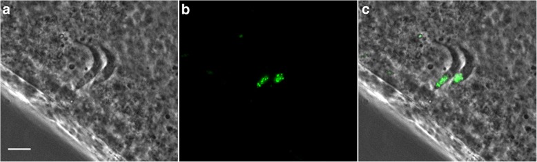
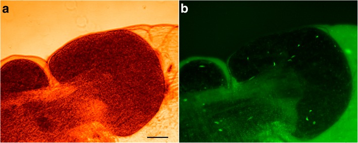
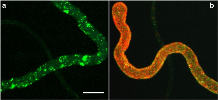
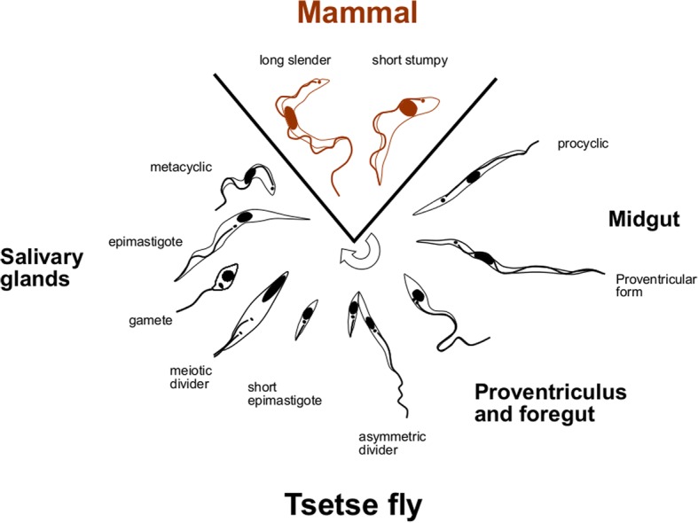
The discovery and development of fluorescent proteins for the investigation of living cells and whole organisms has been a major advance in biomedical research. This approach was quickly exploited by parasitologists, particularly those studying single-celled protists. Here we describe some of our experiments to illustrate how fluorescent proteins have helped to reveal what trypanosomes get up to inside the tsetse fly. Fluorescent proteins turned the tsetse fly from a "black box" into a bright showcase to track trypanosome migration and development within the insect. Crosses of genetically modified red and green fluorescent trypanosomes produced yellow fluorescent hybrids and established the "when" and "where" of trypanosome sexual reproduction inside the fly. Fluorescent-tagging endogenous proteins enabled us to identify the meiotic division stage and gametes inside the salivary glands of the fly and thus elucidate the mechanism of sexual reproduction in trypanosomes. Without fluorescent proteins we would still be in the "dark ages" of understanding what trypanosomes get up to inside the tsetse fly.
Keywords: Fluorescent proteins; Gametes; Glossina; Meiosis; Sexual reproduction; Trypanosoma brucei; Tsetse.
Conflict of interest statement
Ethics approval and consent to participate
Not applicable.
Consent for publication
Not applicable.
Competing interests
The authors declare that they have no competing interests.
Publisher’s Note
Springer Nature remains neutral with regard to jurisdictional claims in published maps and institutional affiliations.
Figures


References
-
- Smyth JD. Introduction to Animal Parasitology. 3. Cambridge: Cambridge University Press; 1994.
Publication types
MeSH terms
Grants and funding
LinkOut - more resources
Full Text Sources
