. 2019 Jan 4;18(1):2.
doi: 10.1186/s12943-018-0929-3.

Low levels of tumour suppressor miR-655 in plasma contribute to lymphatic progression and poor outcomes in oesophageal squamous cell carcinoma

Affiliations

Low levels of tumour suppressor miR-655 in plasma contribute to lymphatic progression and poor outcomes in oesophageal squamous cell carcinoma

Jun Kiuchi et al. Mol Cancer. .

Abstract

Recent studies identified that low levels of tumour suppressor microRNAs (miRNAs) in plasma/serum relate to tumour progression and poor outcomes in cancers. We selected six candidates (miR-126, 133b, 143, 203, 338-3p, 655) of tumour suppressor miRNAs in oesophageal squamous cell carcinoma (ESCC) by a systematic review of NCBI database. Of these, miR-655 levels were significantly down-regulated in plasma of ESCC patients compared to healthy volunteers by test- and validation-scale analyses. Low levels of plasma miR-655 were significantly associated with lymphatic invasion, lymph node metastasis and advanced stage. Univariate and multivariate analysis revealed that the low level of plasma miR-655 was an independent risk factor of lymphatic progression and a poor prognostic factor. Overexpression of miR-655 in ESCC cells inhibited cell proliferation, migration, invasion and epithelial-mesenchymal transition. Increased plasma miR-655 levels by the subcutaneous injection significantly inhibited lymph node metastasis in mice. Low levels of miR-655 in plasma relate to lymphatic progression and poor outcomes, and the restoration of the plasma miR-655 levels might inhibit tumour and lymphatic progression in ESCC.

Keywords: Biomarker; Lymph node metastasis; Mouse model; Oesophageal squamous cell carcinoma; Plasma microRNA; Therapeutic agent.


Conflict of interest statement

Ethics approval

All experimental methods were carried out in accordance with relevant guidelines and regulations, such as the Declaration of Helsinki. Written informed consent was obtained from all patients to use their tissue specimens and blood samples. This study was approved by the institutional review boards of Kyoto Prefectural University of Medicine (ERB-C-319-1). The animal protocol was approved by the Institutional Animal Care and Use Committee of Kyoto Prefectural University of Medicine, and all experiments were conducted strictly in accordance to the National Institute of Health Guide for Care and Use of Laboratory Animals.

Consent for publication

Not applicable in this study.

Competing interests

The authors declare that they have no competing interests.

Publisher’s Note

Springer Nature remains neutral with regard to jurisdictional claims in published maps and institutional affiliations.

Figures

Fig. 1
Fig. 1
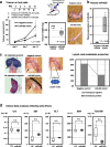
Large-scale analysis of the miR-655 plasma levels in ESCC patients and healthy volunteers, and investigation whether miR-655 would suppress tumour progression and EMT in ESCC cells. a We confirmed that the plasma levels of miR-655 was significantly lower in ESCC patients than in healthy volunteers (P < 0.001). b Decreased plasma miR-655 was associated with poor prognostic outcomes in ESCC patients (P = 0.028). c Cell proliferation analysis by miR-655 overexpression. Proliferation was significantly suppressed in KYSE790 and TE5 cells transfected with the miR-655 mimic compared with cells transfected with the negative control mimic. The FACS analysis demonstrated that transfection of KYSE790 and TE5 cells with the miR-655 mimic resulted in an accumulation of cells in the G1-S phase compared with transfection with the control miRNA mimic. In the protein analysis, overexpression of miR-655 induced the production of p21 and PTEN proteins at 72 h after transfecting the cells with the miR-655 mimic. d Trans-well migration and invasion assays demonstrated that miR-655 suppressed the ability of ESCC cells to migrate and invade. e Overexpression of miR-655 induced a significant morphological change, increased the expression of E-cadherin protein, and reduced the expression of Vimentin, Snail, and ZEB1 proteins
Fig. 2
Fig. 2
Restoration of the miR-655 plasma levels could suppress tumour growth and lymph node metastasis in vivo. a Investigation into whether miR-655 could suppress tumour growth in vivo. The popliteal lymph node metastasis model was used to assess the therapeutic effect of miR-655 for lymph node metastasis. Seven days after the injection of KYSE790 cells into the footpads, tumour development at the injection site was visually confirmed every 7 days. The miR-655 or negative control mimic with atelocollagen was subcutaneously injected into the ventral surface of the lower flank region, which is far from the region injected with cancer cells, and these treatments continued weekly for three weeks after the initial treatment. The treatment with miR-655 mimic significantly suppressed tumour growth on the footpads compared with the negative control mimic. b Comparison of the plasma miR-655 levels between mice treated with miR-655 mimic and treated with negative control mimic. The plasma levels of miR-655 were significantly higher in mice treated with miR-655 mimic than in mice treated with negative control mimic. c Investigation into whether treatment with miR-655 mimic could suppress lymph node metastasis in vivo. To diagnose the lymph node metastasis, we resected the popliteal lymph node on the affected side and sectioned each lymph node into three slices. Lymph node metastasis was diagnosed by hematoxylin–eosin staining. The proportion of lymph node metastasis was significantly lower in mice treated with miR-655 mimic than in mice treated with negative control mimic. d Blood data analyses reflecting potential side effects following miR-655 treatment. There were no side effects in blood-based parameters of organ disorders such as creatinine (Cre), aspartate aminotransferase (AST), alanine aminotransferase (ALT), amylase (AMY), and total bilirubin (T-Bil)

Similar articles

Cited by

References

    1. He L, Thomson JM, Hemann MT, Hernando-Monge E, Mu D, Goodson S, Powers S, Cordon-Cardo C, Lowe SW, Hannon GJ, Hammond SM. A microRNA polycistron as a potential human oncogene. Nature. 2005;435:828–833. doi: 10.1038/nature03552. - DOI - PMC - PubMed
    1. Calin GA, Croce CM. MicroRNA signatures in human cancers. Nat Rev Cancer. 2006;6:857–866. doi: 10.1038/nrc1997. - DOI - PubMed
    1. Mitchell PS, Parkin RK, Kroh EM, Fritz BR, Wyman SK, Pogosova-Agadjanyan EL, Peterson A, Noteboom J, O'Briant KC, Allen A, et al. Circulating microRNAs as stable blood-based markers for cancer detection. Proc Natl Acad Sci U S A. 2008;105:10513–10518. doi: 10.1073/pnas.0804549105. - DOI - PMC - PubMed
    1. Tsujiura M, Ichikawa D, Komatsu S, Shiozaki A, Takeshita H, Kosuga T, Konishi H, Morimura R, Deguchi K, Fujiwara H, et al. Circulating microRNAs in plasma of patients with gastric cancers. Br J Cancer. 2010;102:1174–1179. doi: 10.1038/sj.bjc.6605608. - DOI - PMC - PubMed
    1. Ichikawa D, Komatsu S, Konishi H, Otsuji E. Circulating microRNA in digestive tract cancers. Gastroenterology. 2012;142:1074–1078.e1071. doi: 10.1053/j.gastro.2012.03.008. - DOI - PubMed