. 2019 Mar 8;294(10):3454-3463.
doi: 10.1074/jbc.RA118.004571. Epub 2019 Jan 4.

An alternative N-terminal fold of the intestine-specific annexin A13a induces dimerization and regulates membrane-binding

Affiliations

An alternative N-terminal fold of the intestine-specific annexin A13a induces dimerization and regulates membrane-binding

Kathryn M McCulloch et al. J Biol Chem. .

Abstract

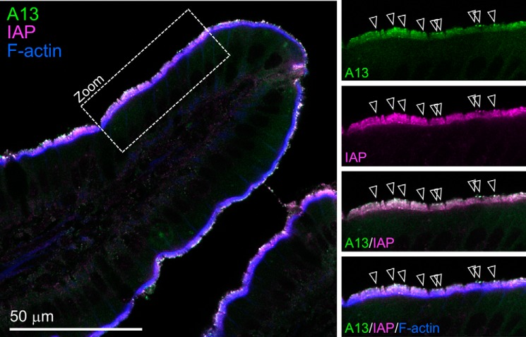
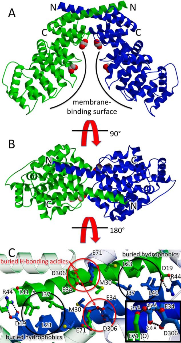
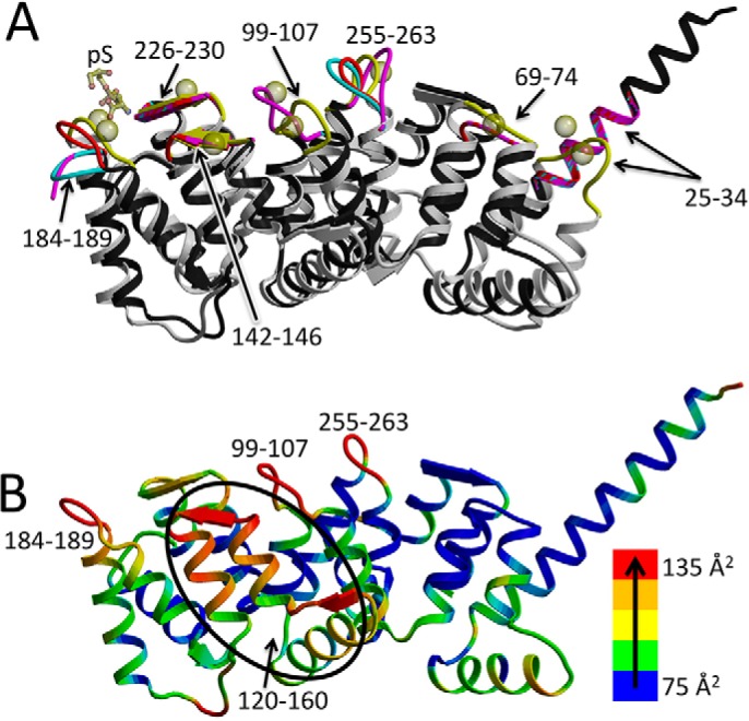
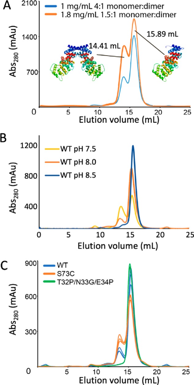
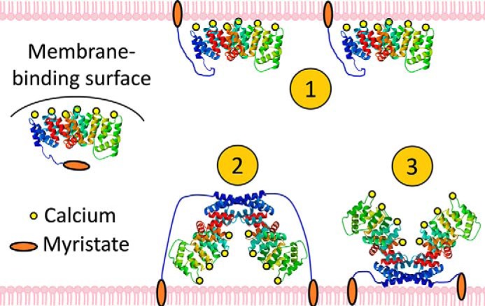
Annexin proteins function as Ca2+-dependent regulators of membrane trafficking and repair that may also modulate membrane curvature. Here, using high-resolution confocal imaging, we report that the intestine-specific annexin A13 (ANX A13) localizes to the tips of intestinal microvilli and determined the crystal structure of the ANX A13a isoform to 2.6 Å resolution. The structure revealed that the N terminus exhibits an alternative fold that converts the first two helices and the associated helix-loop-helix motif into a continuous α-helix, as stabilized by a domain-swapped dimer. We also found that the dimer is present in solution and partially occludes the membrane-binding surfaces of annexin, suggesting that dimerization may function as a means for regulating membrane binding. Accordingly, as revealed by in vitro binding and cellular localization assays, ANX A13a variants that favor a monomeric state exhibited increased membrane association relative to variants that favor the dimeric form. Together, our findings support a mechanism for how the association of the ANX A13a isoform with the membrane is regulated.

Keywords: annexin; calcium regulation; intestinal microvilli; membrane curvature; membrane fusion; oligomerization; protein folding; protein structure; structure-function.


Conflict of interest statement

The authors declare that they have no conflicts of interest with the contents of this article

Figures

Figure 1.
Figure 1.
Localization of ANX A13 in mouse intestinal tissue. Laser scanning confocal image of mouse small intestine tissue section labeled with anti-ANX A13 (green); anti-IAP, a protein that is enriched in luminal vesicles (magenta); and phalloidin to highlight F-actin (blue). Panels at right show higher magnification views from region marked Zoom. Arrowheads in merge panel mark puncta with high levels of ANX A13 and IAP colocalization.
Figure 2.
Figure 2.
Domain swapped dimer of ANX A13a. In the ribbon diagram, one protomer is colored blue, one protomer is colored green, and the N and C termini are labeled. Putative ethylene glycol molecules from the cryoprotectant that are bound to the predicted lipid sites are shown as space-filling models. The two views are separated by 90°. A, view of the domain-swapped dimer perpendicular to the molecular 2-fold axis. The membrane-binding surface is marked with a curved line. Access to the predicted membrane-binding surface is occluded by dimer formation. B, rotation of the view by 90° shows the domain-swapped dimer down the molecular 2-fold axis. C, interactions of the extended helices. The view is from the protein interior such to highlight buried side chains. This identifies hydrophobic interactions (black circles) and six unusual hydrogen bonds between acidic residues (three in each of the red circles). The acidic hydrogen bonding would suggest that Glu-34 and Asp-306 share at least two protons. The inset highlights these hydrogen-bonds; the view of the inset is rotated 30° around the y axis as compared with the rest of the panel.
Figure 3.
Figure 3.
Ca2+-binding sites of ANX A13a. A, overlay of the structure of the three molecules of ANX A13a in each asymmetric unit (black with red, cyan, and magenta Ca2+-binding loops) with ANX A5 (Protein Data Bank entry 1A8A (5); gray backbone with yellow loops) bound of Ca2+ (yellow spheres) and phosphoserine (yellow sticks). The positions of the Ca2+-binding loops are marked and are among the regions of greater structural dissimilarity. B, relative temperature factors of ANX A13a. The structure of ANX A13a (A chain) is colored according to temperature factor, with the lowest temperature factor in blue and ramping via green, yellow, and orange to the highest temperature factor in red. Sequence regions containing the highest temperature factors are labeled and correlate with the Ca2+-binding loops.
Figure 4.
Figure 4.
Monomer–dimer equilibrium of WT and variant ANX A13a in solution. A, evaluation of the elution profile of WT ANX A13a at 1 and 1.8 mg/ml. WT ANX A13a eluted as a doublet, with calculated molecular masses of 37.9 ± 3.7 and 74.5 ± 6.4 kDa. These values are consistent with the theoretical molecular masses of an ANX 13a monomer (35.4 kDa) and dimer (70.8 kDa). At a concentration of 1 mg/ml (blue trace), the monomer:dimer ratio is 4:1, whereas increasing the concentration to 1.8 mg/ml (orange trace) increases the percentage of the dimer such that the monomer:dimer ratio is now 1.5:1. Analysis was done in triplicate with a representative replicate shown. B, evaluation of the elution profile of 1 mg/ml wild-type ANX A13a at pH 7.0, 7.5, and 8.5. The peak corresponding to the ANX A13a dimer is reduced as pH increases. Analysis was performed in triplicate with a representative chromatogram shown. C, comparison of dimerization propensity of purified WT and variant ANX A13a at a concentration of 0.5 mg/ml. WT ANX A13a (blue trace) exhibits a distinct monomer:dimer distribution. The ANX A13aT32P/N33G/E34P (green trace) designed monomer exhibits no detectable dimer. The ANX A13aS73C (orange trace) designed dimer exhibits an increase in dimerization over WT but is not fully converted to the dimeric form under the conditions tested. Analysis was performed in triplicate with each replicate shown.
Figure 5.
Figure 5.
SDS-PAGE analysis for WT and variant ANX A13a. The numbers refer to the elution volume from the size-exclusion column shown in Fig. 4. Both the monomer and dimer peak migrate at the same molecular weight. For the S73C variant, fractions were evaluated under both reducing (unlabeled) and non-reducing (NR) conditions, showing that disulfide is resistant to reduction.
Figure 6.
Figure 6.
Hypotheses for membrane binding by ANX A13a. The canonical membrane-binding surface of ANX A13a is shielded by the domain-swapped dimer, but constitutive membrane association is promoted by the N-terminal myristoylation. Multiple membrane-interacting modes could be envisioned: 1) ANX A13a disassociates into monomers and interacts with the membrane via both the myristoylation and the membrane-remodeling surface; 2) the dimer interacts with the membrane weakly via the canonical membrane-binding surface and strongly via the myristoylation; and 3) the dimer remains membrane-associated via myristoylation, but the canonical membrane-remodeling surface is unable to interact with membranes in the oligomer.
Figure 7.
Figure 7.
Ca2+-dependent binding of ANX A13a to phospholipids. Phospholipids with varied compositions (100% DOPC, 70% PC/30% PA, 70% PC/30% PS) were incubated with purified WT ANX A13a, the designed monomer (ANX A13aT32P/N33G/E34P), and the enhanced dimer (ANX A13aS73C) at a ratio of 500:1 and in the presence or absence of 0.5 mm CaCl2 (± Ca2+). Samples were centrifuged at 100,000 × g, and the resultant supernatant (S) and pellet (P) were separated by SDS-PAGE. The assay was performed in triplicate, with a representative gel shown.
Figure 8.
Figure 8.
Localization of ANX A13a variants expressed in LS174T-W4 cells. A, single plane confocal images of individual LS174T-W4 cells expressing EGPF-tagged ANX A13a, A13aG2A (nonmyristoylated), ANX A13aT32P/N33G/E34P (designed monomer), and A13aS73C (enhanced dimer). Higher intensities are encoded with warmer colors. WT ANX A13a is robustly localized at the cell surface, whereas nonmyristoylated ANX A13a (ANX A13aG2A) is distinctly localized in the cytoplasm. Expression of the designed monomeric form of ANX A13a (ANX A13T32P/N33G/E34P) results in a subtle decrease in surface levels compared with WT ANX A13a. The enhanced dimeric form of ANX A13a exhibits increased cytoplasm localization relative to WT ANX A13a. All boxes are 25 × 25 μm. B, peripheral:cytoplasmic intensity ratios derived from 11–13 cells representing two biological replicates. Pair-wise p values are listed in Table 3.

References

    1. Creutz C. E., Pazoles C. J., and Pollard H. B. (1978) Identification and purification of an adrenal medullary protein (synexin) that causes calcium-dependent aggregation of isolated chromaffin granules. J. Biol. Chem. 253, 2858–2866 - PubMed
    1. Gerke V., Creutz C. E., and Moss S. E. (2005) Annexins: linking Ca2+ signalling to membrane dynamics. Nat. Rev. Mol. Cell Biol. 6, 449–461 10.1038/nrn1671, 10.1038/nrm1661 - DOI - PubMed
    1. Lizarbe M. A., Barrasa J. I., Olmo N., Gavilanes F., and Turnay J. (2013) Annexin–phospholipid interactions: functional implications. Int. J. Mol. Sci. 14, 2652–2683 10.3390/ijms14022652 - DOI - PMC - PubMed
    1. Huber R., Berendes R., Burger A., Luecke H., and Karshikov A. (1992) Annexin V-crystal structure and its implications on function. Behring Inst. Mitt. 107–125 - PubMed
    1. Swairjo M. A., Concha N. O., Kaetzel M. A., Dedman J. R., and Seaton B. A. (1995) Ca2+-bridging mechanism and phospholipid head group recognition in the membrane-binding protein annexin V. Nat. Struct. Biol. 2, 968–974 10.1038/nsb1195-968 - DOI - PubMed

Publication types

LinkOut - more resources