

Teaching Benign Skin Lesions as a Strategy to Improve the Triage Amalgamated Dermoscopic Algorithm (TADA)
- PMID: 30610147
- PMCID: PMC8039813
- DOI: 10.3122/jabfm.2019.01.180049
Teaching Benign Skin Lesions as a Strategy to Improve the Triage Amalgamated Dermoscopic Algorithm (TADA)
Abstract
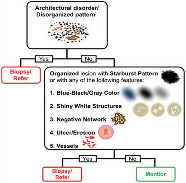
Introduction: Dermoscopy aids family physicians (FPs) in skin cancer detection. The triage amalgamated dermoscopic algorithm (TADA) was created to simplify the dermoscopic evaluation of a skin growth. The purpose of this image-based study was to evaluate the effect of teaching the clinical and dermoscopic features of benign skin lesions on the diagnostic accuracy of skin cancer identification using TADA. We also sought to determine the best method to teach benign neoplasms.
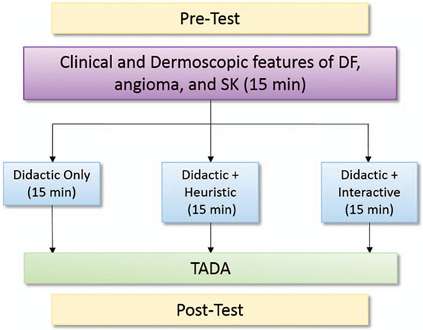
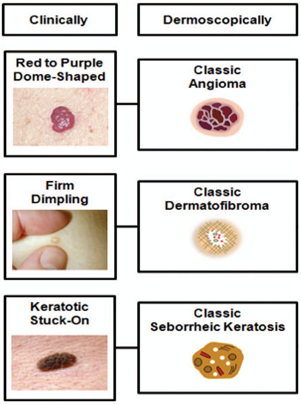
Methods: In this cross-sectional study of an educational intervention, FPs participated in dermoscopy training. Participants were divided into 3 groups for teaching of common benign neoplasms (dermatofibroma, angioma, and seborrheic keratosis/lentigo): didactic + interactive, didactic + heuristic, and didactic. For each group, the benign teaching was followed by skin cancer identification training with TADA. All participants took a 30 image pre-test and 30 image post-test.
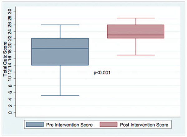
Results: Fifty-nine participants completed the study. The mean preintervention score (out of 30 correct responses) was 17.9 (SD, 4.5) and increased to 23.5 (SD, 3.0) on the postintervention evaluation (P < .001). Sensitivity for skin cancer increased from 62.5% to 88.1% following the intervention. Postintervention specificity for skin cancer was 87.8%. Sensitivity and specificity increased following the intervention for all 3 types of benign neoplasms. Diagnostic accuracy was not impacted by the method of benign teaching.
Conclusion: Short dermoscopy training sessions with dedicated time for benign growths followed by TADA training for malignant growths are an effective means of teaching FPs dermoscopy and result in a high sensitivity and specificity for the identification of benign and malignant skin neoplasms.
Keywords: Cross-Sectional Studies; Dermatofibroma; Dermoscopy; Family Physicians; Seborrheic Keratosis; Skin Cancer.
© Copyright 2019 by the American Board of Family Medicine.
Conflict of interest statement
Conflict of interest: Dr. Marghoob has served as a consultant for 3GEN, Canfield, and Heine. However, he did not receive support from them for any portion of this research or manuscript. He has received honoraria from 3GEN and is friends with employees at 3GEN and Canfield. He has intellectual passion for dermoscopy. The other authors have no conflicts of interest to disclose.
Figures

Similar articles
-
A Clinical Aid for Detecting Skin Cancer: The Triage Amalgamated Dermoscopic Algorithm (TADA).J Am Board Fam Med. 2016 Nov 12;29(6):694-701. doi: 10.3122/jabfm.2016.06.160079. J Am Board Fam Med. 2016. PMID: 28076252 Free PMC article.
-
Dermoscopy Training Effect on Diagnostic Accuracy of Skin Lesions in Canadian Family Medicine Physicians Using the Triage Amalgamated Dermoscopic Algorithm.Dermatol Pract Concept. 2020 Apr 3;10(2):e2020035. doi: 10.5826/dpc.1002a35. eCollection 2020. Dermatol Pract Concept. 2020. PMID: 32363097 Free PMC article.
-
Triage amalgamated dermoscopic algorithm (TADA) for skin cancer screening.Dermatol Pract Concept. 2017 Apr 30;7(2):39-46. doi: 10.5826/dpc.0702a09. eCollection 2017 Apr. Dermatol Pract Concept. 2017. PMID: 28515993 Free PMC article.
-
Reflectance confocal microscopy: Diagnostic criteria of common benign and malignant neoplasms, dermoscopic and histopathologic correlates of key confocal criteria, and diagnostic algorithms.J Am Acad Dermatol. 2021 Jan;84(1):17-31. doi: 10.1016/j.jaad.2020.05.154. Epub 2020 Jun 18. J Am Acad Dermatol. 2021. PMID: 32565210 Review.
-
Dermoscopy is useful for the recognition of benign-malignant compound tumours.Br J Dermatol. 2005 Sep;153(3):653-6. doi: 10.1111/j.1365-2133.2005.06717.x. Br J Dermatol. 2005. PMID: 16120160 Review.
Cited by
-
Perspectives on Dermoscopy in the Primary Care Setting.J Am Board Fam Med. 2020 Nov-Dec;33(6):1022-1024. doi: 10.3122/jabfm.2020.06.200238. J Am Board Fam Med. 2020. PMID: 33219084 Free PMC article.
-
Absence of central white patch in dermatofibromas presenting in darker skin.JAAD Case Rep. 2022 Jan 6;21:63-65. doi: 10.1016/j.jdcr.2021.12.023. eCollection 2022 Mar. JAAD Case Rep. 2022. PMID: 35198701 Free PMC article. No abstract available.
-
Impact of Dermoscopy Training for Primary Care Practitioners on Number Needed to Biopsy to Detect Melanoma.PRiMER. 2023 Feb 3;7:276659. doi: 10.22454/PRiMER.2023.276659. eCollection 2023. PRiMER. 2023. PMID: 36845847 Free PMC article.
-
Primary Care Physician Use of Elastic Scattering Spectroscopy on Skin Lesions Suggestive of Skin Cancer.J Prim Care Community Health. 2025 Jan-Dec;16:21501319251344423. doi: 10.1177/21501319251344423. Epub 2025 Jun 5. J Prim Care Community Health. 2025. PMID: 40470593 Free PMC article.
-
Educational Interventions to Support Primary Care Provider Performance of Diagnostic Skin Cancer Examinations: A Systematic Literature Review.J Cancer Educ. 2022 Dec;37(6):1579-1588. doi: 10.1007/s13187-021-02118-8. Epub 2022 Jan 18. J Cancer Educ. 2022. PMID: 35040018 Free PMC article.
References
-
- Society AC. Cancer Facts & Figures 2017. Atlanta (GA): American Cancer Society; 2017.
-
- Kerr O, Walker J, Boohan M. General practitioners opinions regarding the need for training in dermatology at undergraduate and postgraduate levels. Clin Exp Dermatol 2006;31:132–3. - PubMed
-
- Vestergaard M, Macaskill P, Holt P, Menzies S. Dermoscopy compared with naked eye examination for the diagnosis of primary melanoma: a meta-analysis of studies performed in a clinical setting. Br J Dermatol 2008;159:669–76. - PubMed
-
- Wozniak-Rito A, Zalaudek I, Rudnicka L. Dermoscopy of basal cell carcinoma. Clin Exp Dermatol 2018;43:241–7. - PubMed
Publication types
MeSH terms
Grants and funding
LinkOut - more resources
Full Text Sources
Miscellaneous