. 2019 Jan;31(1):68-83.
doi: 10.1105/tpc.18.00478. Epub 2019 Jan 4.

A Remote cis-Regulatory Region Is Required for NIN Expression in the Pericycle to Initiate Nodule Primordium Formation in Medicago truncatula

Affiliations

A Remote cis-Regulatory Region Is Required for NIN Expression in the Pericycle to Initiate Nodule Primordium Formation in Medicago truncatula

Jieyu Liu et al. Plant Cell. 2019 Jan.

Abstract

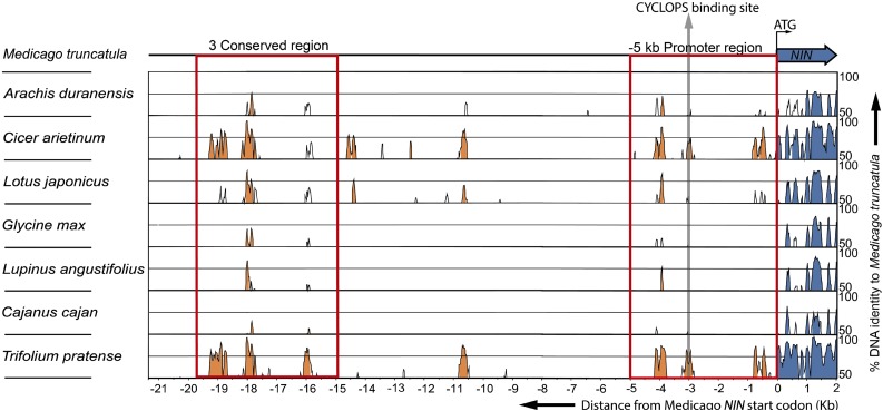
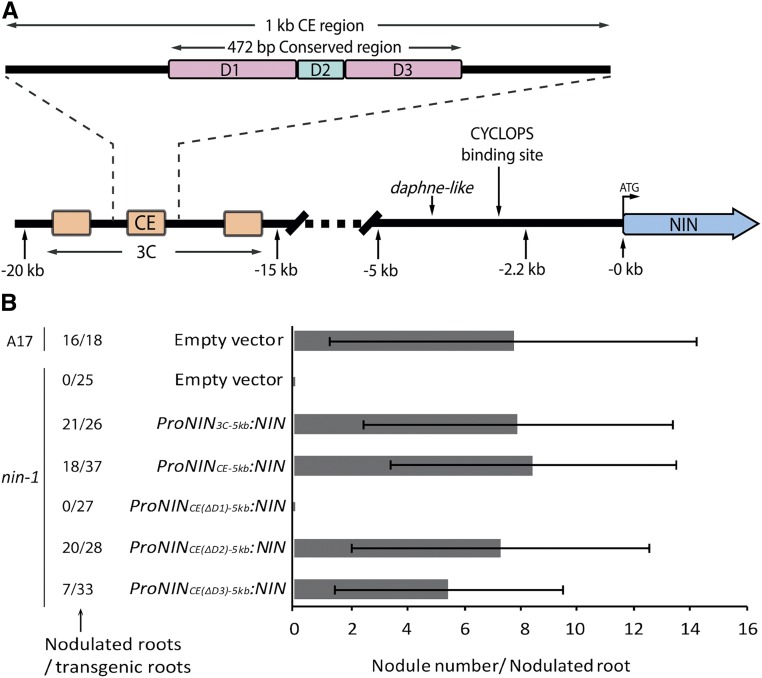
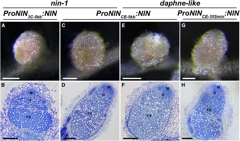
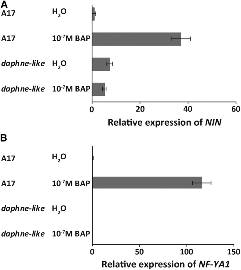
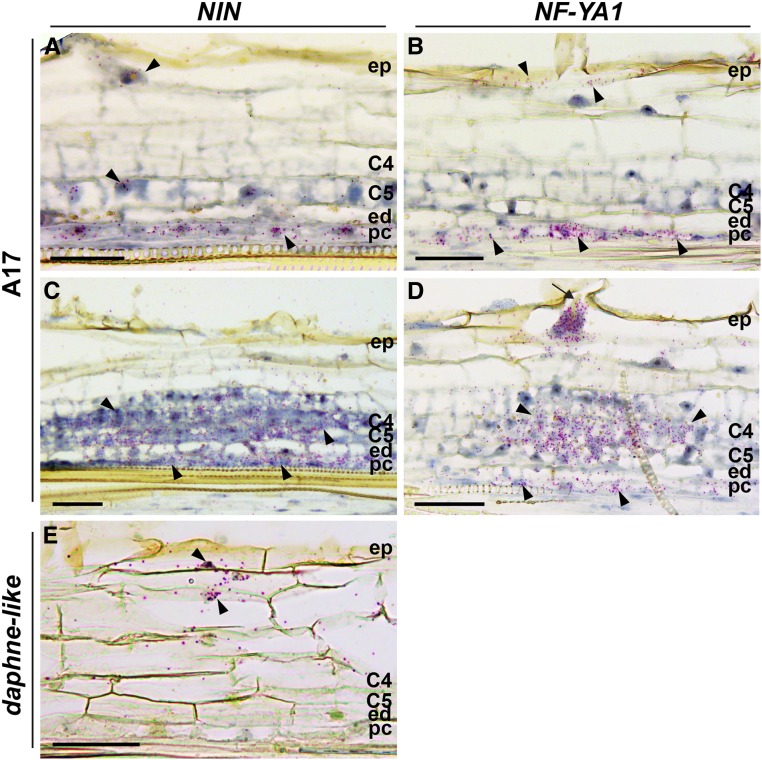
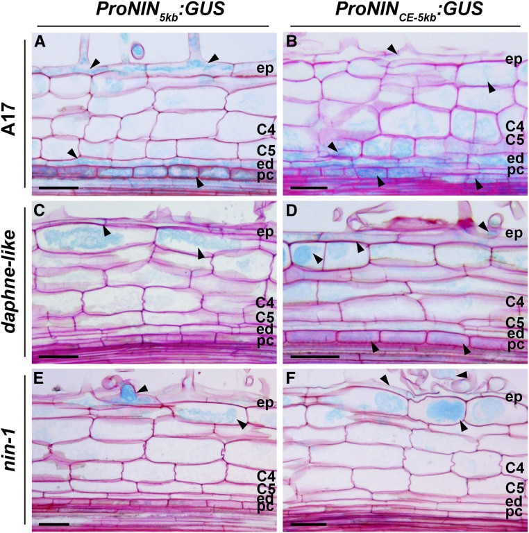
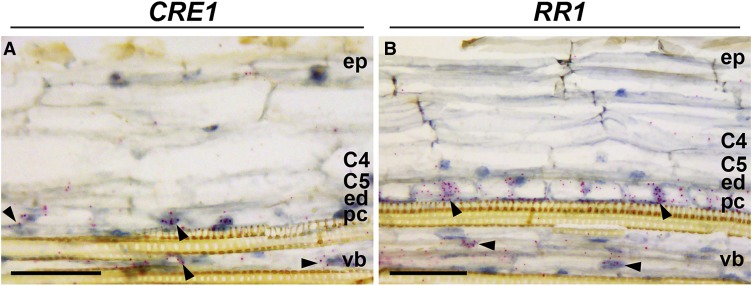
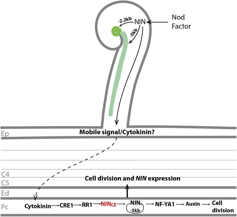
The legume-rhizobium symbiosis results in nitrogen-fixing root nodules, and their formation involves both intracellular infection initiated in the epidermis and nodule organogenesis initiated in inner root cell layers. NODULE INCEPTION (NIN) is a nodule-specific transcription factor essential for both processes. These NIN-regulated processes occur at different times and locations in the root, demonstrating a complex pattern of spatiotemporal regulation. We show that regulatory sequences sufficient for the epidermal infection process are located within a 5 kb region directly upstream of the NIN start codon in Medicago truncatula Furthermore, we identify a remote upstream cis-regulatory region required for the expression of NIN in the pericycle, and we show that this region is essential for nodule organogenesis. This region contains putative cytokinin response elements and is conserved in eight more legume species. Both the cytokinin receptor 1, which is essential for nodule primordium formation, and the B-type response regulator RR1 are expressed in the pericycle in the susceptible zone of the uninoculated root. This, together with the identification of the cytokinin-responsive elements in the NIN promoter, strongly suggests that NIN expression is initially triggered by cytokinin signaling in the pericycle to initiate nodule primordium formation.


Figures

Figure 1.
Figure 1.
In the Medicago nin Mutant daphne-like, Infection and Nodule Organogenesis Are Uncoupled. (A) to (D) Images of wild-type and mutant roots. These transmitted light stereomacroscopy images (A) and (C) and corresponding green fluorescence stereomacroscopy images (B) and (D) were taken at 3 wpi. Roots of daphne-like (FN8113) mutant plants ([C] and [D]) have an excessive number of infection threads in comparison with wild-type A17 roots ([A] and [B]). Bars = 2 mm. (E) Quantification of infection thread number. The number of infection threads per root was counted at 2 wpi in both A17 roots (n = 12) and FN8113 roots (n = 12). Data are means ± sd. (F) and (G) Infection thread formation in mutant and wild-type roots. These confocal images of roots stained with propidium iodide at 1 wpi show that a bacterial colony (arrowheads) is formed inside a daphne-like curled root hair and an infection thread (arrows) is initiated (F) like in a wild-type root hair (G). S. meliloti containing constitutively expressed GFP was used as inoculum. Bars = 10 µm. (H) Longitudinal plastic section of daphne-like root at 3 wpi. The section stained with toluidine blue displays an infection thread (arrows). The infection threads in a mutant can occasionally reach cortical cell layers and induce some cell divisions (arrowheads). C4 and C5, cortical cell layers 4 and 5; ed, endodermis; ep, epidermis; pc, pericycle. Bar = 50 μm. (I) Schematic representation of the chromosome translocation at the NIN locus in the daphne-like mutant. The strikethrough indicates a 15 bp deleted sequence.
Figure 2.
Figure 2.
The Infection Process in Medicago nin-1 Roots Is Partially Rescued by Introducing ProNIN5kb:NIN or ProNIN2.2kb:NIN. (A) to (D) Phenotype of nin-1 roots transformed with ProNIN5kb:NIN at 4 wpi with S. meliloti constitutively expressing GFP. The transmitted light microscopy image of a longitudinal plastic section of a transgenic root stained with toluidine blue displays infection threads (arrow) that occasionally can reach cortical cell layers (A). The transmitted light stereomacroscopy image (B) and corresponding green fluorescence stereomacroscopy image (C) show excessive infection thread formation in transgenic roots. The confocal image of a transgenic root stained with propidium iodide shows an infection thread initiated in the curled root hair (D). Bars = 50 µm (A), 2 mm ([B] and [C]), and 10 µm (D). (E) to (G) Phenotype of nin-1 roots transformed with ProNIN2.2kb:NIN at 4 wpi with S. meliloti constitutively expressing GFP. The transmitted light stereomacroscopy image (E) and corresponding green fluorescence stereomacroscopy image (F) display numerous curled root hairs with bacterial colonies in transgenic roots. In the confocal image of a transgenic root stained with propidium iodide (G), colonies are formed inside the chamber of the root hair curl, but an infection thread does not develop. Bars = 2 mm ([E] and [F]) and 10 µm (G). (H) to (J) Phenotype of nin-1 roots transformed with empty vector at 4 wpi with S. meliloti constitutively expressing GFP. The transmitted light stereomacroscopy image (H) and corresponding green fluorescence stereomacroscopy image (I) show that the transgenic root forms neither infection threads nor bacterial colonies. A transgenic root stained with propidium iodide shows excessive root hair curling (J). Bars = 2 mm ([H] and [I]) and 10 µm (J). (K) to (M) Phenotype of nin-1 roots transformed with ProNIN5kb(Δcyclops):NIN at 4 wpi with S. meliloti constitutively expressing GFP. The transmitted light stereomacroscopy image (K) and corresponding green fluorescence stereomacroscopy image (L) display many bacterial colonies in transgenic root hairs. The confocal image of a transgenic root stained with propidium iodide shows that a colony is formed inside the chamber of the root hair curl but an infection thread is not initiated (M). Bars = 2 mm ([K] and [L]) and 10 µm (M).
Figure 3.
Figure 3.
mVISTA Alignment of Genomic DNA Sequences Including 2 kb from the Start Codon of NIN and 5′-Upstream Regions from Eight Legume Species. Peaks indicate the level of identity with Medicago on a scale of 50 to 100%. Identities lower than 50% were not scored. The figure shows 2 kb of each NIN sequence downstream of the start codon (blue) and the 5′ upstream DNA sequence (orange). The red rectangle on the right next to the NIN coding sequence indicates the –5 kb promoter region. The red rectangle on the left indicates the 3C. The gray arrow shows the putative CYCLOPS binding site.
Figure 4.
Figure 4.
The CE Region in the NIN Promoter Is Essential for Nodule Organogenesis. (A) Schematic illustration of the Medicago NIN promoter. The 3C region (orange) was identified among the eight legume species studied here. The second 3C region is most conserved, and it includes about 10 putative B-type cytokinin signaling RR binding sites and is named the CE region. The CE region in turn contains a highly conserved 472 bp sequence that was divided into three parts named D1, D2, and D3. D1 and D3 (purple) contain six and three putative cytokinin response elements, respectively, whereas D2 (green) contains a putative AP2 binding site and a single putative cytokinin response element. (B) The number of nodules formed on wild-type (A17) roots transformed with empty vector and nin-1 roots transformed with the constructs carrying NIN driven by different parts of the NIN promoter as indicated. S. meliloti containing constitutively expressed GFP was used as inoculum. Nodule numbers per nodulated root were counted at 4 wpi. Data are means ± sd.
Figure 5.
Figure 5.
Nonnodulating Phenotypes of nin-1 and daphne-like Are Rescued by A. rhizogenes-Mediated Transformation with ProNIN3C-5kb:NIN, ProNINCE-5kb:NIN, and ProNINCE-35Smin:NIN. (A) to (D) Nodules formed on nin-1 roots transformed with the indicated constructs at 4 wpi with S. meliloti. Transmitted light macroscopy images of nodules are shown ([A] and [C]). Nodules are pink due to the presence of leghemoglobin. Longitudinal plastic sections of these nodules stained with toluidine blue display normal zonation ([B] and [D]). FX, fixation zone; IF, infection zone; M, meristem. Bars = 2 mm ([A] and [C]) and 200 µm ([B] and [D]). (E) to (H) Nodules formed on daphne-like roots transformed with the indicated constructs at 4 wpi with S. meliloti. Transmitted light macroscopy images of nodules are shown ([E] and [G]). Longitudinal plastic sections of these nodules stained with toluidine blue display normal zonation ([F] and [H]). Bars = 2 mm ([E] and [G]) and 200 µm ([F] and [H]).
Figure 6.
Figure 6.
NIN and NF-YA1 Expression Cannot Be Induced by Cytokinin Application in daphne-like Mutants. Quantitative real-time RT-PCR shows NIN (A) and NF-YA1 (B) expression in wild-type (A17) and daphne-like roots 16 h after application of 10−7 M benzylaminopurine (BAP) or water. Data are means ± se of three biological replicates.
Figure 7.
Figure 7.
NIN and NF-YA1 Expression Patterns in Medicago Wild-Type (A17) Nodule Primordia and daphne-like Mutant. (A) to (D) RNA in situ localization of NIN ([A] and [C]) and NF-YA1 ([B] and [D]) in nodule primordia at 2 dpi ([A] and [B]) and at 3 dpi ([C] and [D]). The arrow indicates an infection thread. (E) RNA in situ localization of NIN in roots of the daphne-like mutant at 2 dpi. Hybridization signals are visible as red dots (arrowheads). C4 and C5, cortical cell layers 4 and 5; ed, endodermis; ep, epidermis; pc, pericycle. Bars = 50 µm.
Figure 8.
Figure 8.
The CE Region Is Required for Rhizobium-Induced NIN Expression in the Pericycle. Tissue-specific ProNIN5kb:GUS and ProNINCE-5kb:GUS expression patterns are shown in the wild type and nin mutants at 2 dpi. Arrowheads indicate GUS expression (light blue) in wild-type ([A] and [B]), in daphne-like ([C] and [D]), and in nin-1 ([E] and [F]) roots. C4 and C5, cortical cell layers 4 and 5; ed, endodermis; ep, epidermis; pc, pericycle. Bars = 50 µm.
Figure 9.
Figure 9.
CRE1 and RR1 Are Expressed in the Pericycle of Uninoculated Roots. RNA in situ localization of CRE1 (A) and RR1 (B) is shown in the susceptible zone of uninoculated roots. For in situ hybridization, root tips of ∼1 cm of 4-d-old seedlings were used. Hybridization signals are visible as red dots (arrowheads). C4 and C5, cortical cell layers 4 and 5; ed, endodermis; ep, epidermis; pc, pericycle; vb, vascular bundle. Bars = 50 µm.
Figure 10.
Figure 10.
Proposed Model of NIN Function during Initiation of Nodule Primordia. After perception of the Nod factor, NIN expression is induced in the epidermis. The –5 kb regulatory region of the NIN promoter is sufficient for both tight root hair curling and infection thread formation. By contrast, expression driven by the –2.2 kb region is sufficient only for the tight root hair curling and formation of bacterial colonies inside the curl. A mobile signal is generated in the epidermis in a NIN-dependent manner, and this signal translocates to the pericycle. Whether or not this mobile signal is cytokinin or an unknown signal, it causes cytokinin accumulation in the inner root cell layers. The CRE1 receptor in the pericycle perceives cytokinin and activates the B-type RR1, which further activates NIN expression. NIN directly or indirectly regulates its own expression via a positive feedback loop, and the –5 kb promoter region is sufficient for this feedback regulation. NIN directly activates NF-YA1 expression and stimulates further cell divisions. Later, the NIN-induced response in pericycle contributes to cell division and NIN expression in the endodermis and cortical cells. C4 and C5, cortical cell layers 4 and 5; Ed, endodermis; Ep, epidermis; Pc, pericycle.

Comment in

Similar articles

Cited by

References

    1. Altschul S.F., Gish W., Miller W., Myers E.W., Lipman D.J. (1990). Basic local alignment search tool. J. Mol. Biol. 215: 403–410. - PubMed
    1. Andriankaja A., Boisson-Dernier A., Frances L., Sauviac L., Jauneau A., Barker D.G., de Carvalho-Niebel F. (2007). AP2-ERF transcription factors mediate Nod factor dependent Mt ENOD11 activation in root hairs via a novel cis-regulatory motif. Plant Cell 19: 2866–2885. - PMC - PubMed
    1. Benfey P.N., Chua N.-H. (1990). The cauliflower mosaic virus 35S promoter: Combinatorial regulation of transcription in plants. Science 250: 959–966. - PubMed
    1. Bertioli D.J., et al. (2016). The genome sequences of Arachis duranensis and Arachis ipaensis, the diploid ancestors of cultivated peanut. Nat. Genet. 48: 438–446. - PubMed
    1. Boivin S., Kazmierczak T., Brault M., Wen J., Gamas P., Mysore K.S., Frugier F. (2016). Different cytokinin histidine kinase receptors regulate nodule initiation as well as later nodule developmental stages in Medicago truncatula. Plant Cell Environ. 39: 2198–2209. - PubMed

Publication types

MeSH terms

LinkOut - more resources