. 2019 Jan;31(1):172-188.
doi: 10.1105/tpc.18.00382. Epub 2019 Jan 4.

Binding of the Magnaporthe oryzae Chitinase MoChia1 by a Rice Tetratricopeptide Repeat Protein Allows Free Chitin to Trigger Immune Responses

Affiliations

Binding of the Magnaporthe oryzae Chitinase MoChia1 by a Rice Tetratricopeptide Repeat Protein Allows Free Chitin to Trigger Immune Responses

Chao Yang et al. Plant Cell. 2019 Jan.

Abstract

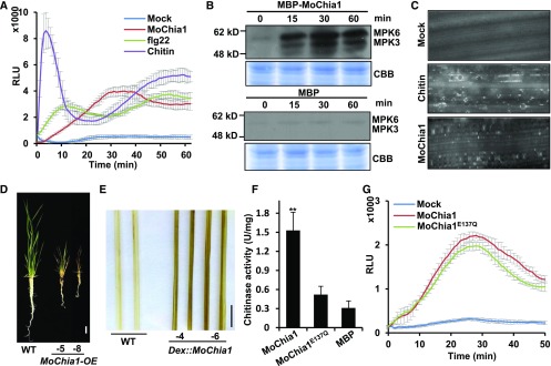
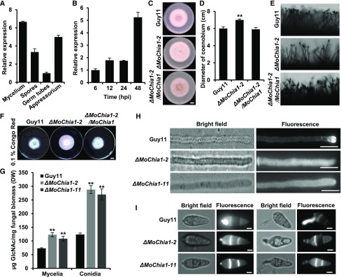
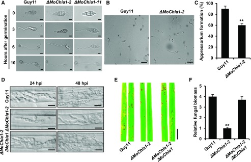
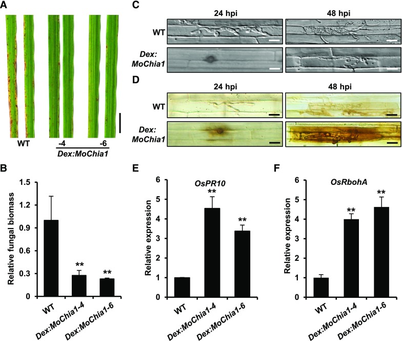
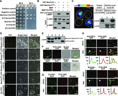
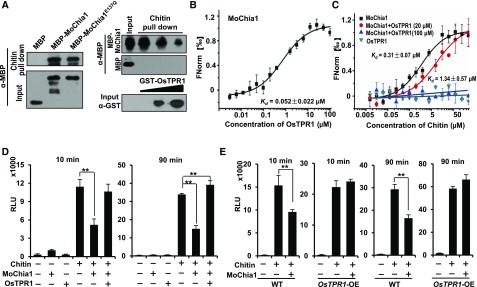
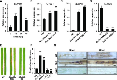
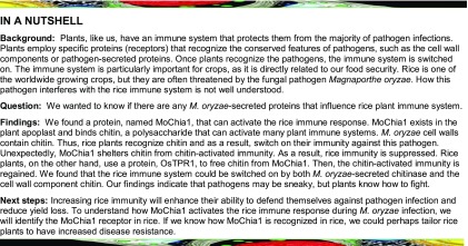
To defend against pathogens, plants have developed complex immune systems, including plasma membrane receptors that recognize pathogen-associated molecular patterns, such as chitin from fungal cell walls, and mount a defense response. Here, we identify a chitinase, MoChia1 (Magnaporthe oryzae chitinase 1), secreted by M. oryzae, a fungal pathogen of rice (Oryza sativa). MoChia1 can trigger plant defense responses, and expression of MoChia1 under an inducible promoter in rice enhances its resistance to M. oryzae MoChia1 is a functional chitinase required for M. oryzae growth and development; knocking out MoChia1 significantly reduced the virulence of the fungus, and we found that MoChia1 binds chitin to suppress the chitin-triggered plant immune response. However, the rice tetratricopeptide repeat protein OsTPR1 interacts with MoChia1 in the rice apoplast. OsTPR1 competitively binds MoChia1, thereby allowing the accumulation of free chitin and re-establishing the immune response. Overexpressing OsTPR1 in rice plants resulted in elevated levels of reactive oxygen species during M. oryzae infection. Our data demonstrate that rice plants not only recognize MoChia1, but also use OsTPR to counteract the function of this fungal chitinase and regain immunity.


Figures

Figure 1.
Figure 1.
MoChia1 Activates Pathogen-Triggered Immune Responses in Rice. (A) MoChia1 activates the ROS burst in rice suspension cells. Recombinant MBP-MoChia1 protein was purified from E. coli, and 2 μg/ml protein was used for the luminol-based ROS burst assay. Flg22 (100 nM) and chitin (0.1 μg/ml) served as positive controls. MBP served as a negative control. Values are means ± sd (n = 4). RLU, relative light units. (B) MoChia1 protein can activate MAP kinase signaling in rice. MoChia1 protein (1 μg/ml) was incubated with rice suspension cells. MBP protein (1 μg/ml) was used as the negative control. Activated MAPKs were detected by immunoblotting with the phospho-p38 MAPK antibody at the indicated times. The corresponding bands are represented for the phosphorylation of mitogen-activated protein kinase 3 and mitogen-activated protein kinase 6. Coomassie brilliant blue (CBB) staining of ribulose-1,5-bis-phosphate carboxylase/oxygenase was used to ensure equal loading in each lane. The experiment was repeated three times with similar results. (C) MoChia1 induces callose deposition in rice. The leaves of 2-week-old rice seedlings were treated with 0.6 μg/ml MoChia1 or 8 nM chitin for 16 h, and then stained using aniline blue. MBP served as the mock treatment. (D) The overexpression of MoChia1 leads to an autoimmune response in rice. MoChia1, driven by the Ubiquitin promoter, was ligated into the pC1390U binary vector and transformed into the rice variety Nipponbare (wild type). Lines 5 and 8 are two representative independent lines. Bar = 1 cm. (E) MoChia1 overexpression leads to ROS accumulation in rice. Two-week-old DEX:MoChia1 plants were treated with 30 μM DEX for 24 h; then the leaves were stained with DAB. Lines 4 and 6 are two independent transgenic lines. Bar = 1 cm. (F) MoChia1 is a functional chitinase, and the Glu137 mutation abolishes its enzymatic activity. The chitinase enzyme activity of the recombinant MBP-MoChia1 protein was analyzed, using colloid chitin as substrate. The reaction of DNS (3,5-dinitrosalicylic acid) and GlcNAc was monitored at OD 565 nm. One unit of chitinase activity was defined as the amount of enzyme required to release 1 μmol of N-acetyl-d-glucosamine per hour. Values are means ± sd (n = 3). **Significant differences from MBP at P < 0.01 (Student’s t test). (G) The enzyme activity of MoChia1 is not required for the ROS burst. Rice suspension cells were used to measure ROS production in a luminol-based assay, using 1 μg/ml protein. MBP was used as a negative control. Values are means ± sd (n = 4).
Figure 2.
Figure 2.
MoChia1 is Required for the Growth and Development of M. oryzae. (A) MoChia1 expression levels in different tissue or developmental stages. MoChia1 expression was investigated using RT-qPCR. The fungi were grown in CM media. Values are means ± sd (n = 3). (B) MoChia1 expression levels during M. oryzae infection in rice. The rice leaves were inoculated with M. oryzae spores at a concentration of 1 × 10⁵ per mL. The leaves were sampled at the indicated time points for RT-qPCR assays. Values are means ± sd (n = 3). (C) MoChia1 affects mycelial growth. The images were taken from the wild-type Guy11 and ΔMoChia1 mutants grown on the CM medium for 2 weeks. Bar = 1 cm. (D) Statistical analysis of the growth rate of mycelia in (C). Values are means ± sd (n = 8). **Significant differences from wild type at P < 0.01 (Student’s t test). (E) ΔMoChia1 mutants produce fewer conidia. Conidia were observed under a light microscope after illumination for 24 h. The experiments were repeated three times with similar results. (F) ΔMoChia1 mutants are less sensitive to cell wall stress. The Guy11, ΔMoChia1 mutants, and complementation strains were grown on complete medium containing 0.1% Congo Red. Photographs were taken after 7 d in culture at 28°C. Bar = 1 cm. (G) ΔMoChia1 mutants display chitin accumulation in the mycelia and conidia. The chitin contents of the Guy11 and ΔMoChia1 mycelia were measured after a 2-d culture at 28°C. Fresh conidia after light induction were used for chitin content measurements. DW, dry weight. Values are means ± sd (n = 3). **Significant differences from Guy11 at P < 0.01 (Student’s t test). (H) and (I) Knocking out MoChia1 leads to abnormal chitin deposition in the mycelia, conidia, and germination tubes. Mycelia were cultured for 14 days at 28°C. Mycelia, fresh conidia, and germinated conidia were used for cell wall staining with calcofluor. The fluorescence was observed under a fluorescence microscope using the DAPI channel. Bar = 10 μm.
Figure 3.
Figure 3.
MoChia1 is Required for M. oryzae Virulence. (A) Germination of conidial spores of the wild type and ΔMoChia1 mutants. Conidia were germinated on glass cover slips. The germination tubes and appressorium development were observed at the indicated time points. ΔMoChia1-2 and ΔMoChia1-11 are two independent mutant strains. Bar = 10 μm. (B) Appressorium formation is largely delayed in the ΔMoChia1 mutants. Appressorium development was observed after a 6-h incubation on a hydrophobic glass surface. Bar = 100 μm. (C) Statistical analysis of appressorium formation in the wild type and ΔMoChia1 mutants observed in (B). Values are means ± sd (n = 3). **Significant differences from the wild type at P < 0.01 (Student’s t test). (D) ΔMoChia1 display reduced virulence on rice. The rice leaf sheath was inoculated with a conidial suspension at a concentration of 1 × 10⁵ conidia per mL in 0.2% Tween 20. The hyphae images were taken at 24 and 48 hpi. Bar = 10 μm. (E) Disease symptoms of rice leaves infected with the wild-type M. oryzae, ΔMoChia1 mutants, and the complementation strains. Conidial suspensions (1 × 10⁵ conidia per mL in 0.2% Tween 20) were sprayed onto the leaf surfaces of 2-week-old seedlings. Images were taken at 7 dpi (days post inoculation). Bar = 1 cm. (F) Relative fungal biomass for (E). The fungal biomass was determined using qPCR of the M. oryzae Pot2 gene against the rice OsUbi1 gene. Values are means ± sd (n = 4). **Significant differences from the wild type at P < 0.01 (Student’s t test).
Figure 4.
Figure 4.
Ectopic Expression of MoChia1 Enhances Rice Resistance to M. oryzae. (A) DEX:MoChia1 plants exhibit enhanced disease resistance to M. oryzae after DEX treatment. Two-week-old DEX:MoChia1 plants were treated with 30 μM DEX applied to the roots. After 24 h, the seedlings were spray-inoculated with conidial suspensions (1 × 10⁵ conidia per mL in 0.2% Tween 20). The images were taken at 5 dpi. Bar = 1 cm. (B) Relative fungal biomass in (A). Values are means ± sd (n = 4). **Significant differences from wild type at P < 0.01 (Student’s t test). (C) The DEX:MoChia1 plants displayed enhanced disease resistance to M. oryzae after DEX treatment. Rice leaf sheaths were inoculated with conidial suspension at a concentration of 1 × 10⁵ conidia per mL in 0.2% Tween 20. The hyphae images were taken at 24 and 48 hpi, respectively. Bar = 10 μm. (D) DEX:MoChia1 plants exhibit enhanced ROS accumulation during M. oryzae infection after DEX treatment. The rice leaf sheath was inoculated with a conidial suspension at a concentration of 1 × 10⁵ conidia per mL in 0.2% Tween 20. The DAB staining was performed at 24 and 48 hpi, respectively. Bar = 10 μm. (E) and (F) OsPR10 and OsRbohA expression was elevated in DEX:MoChia1 plants. Two-week-old wild type and DEX:MoChia1 plants were pretreated with 30 μM DEX. After 24 h, RT-qPCR was used to examine the gene transcription levels in plants. Values are means ± sd (n = 4). **Significant differences from wild type at P < 0.01 (Student’s t test).
Figure 5.
Figure 5.
MoChia1 Interacts with OsTPR1 In Vitro and In Vivo. (A) MoChia1 and MoChia1E137Q interact with OsTPR1 in yeast. AD-MoChia1, AD-MoChia1E137Q, and BD-OsTPR1 plasmids were cotransformed into yeast cells and screened on synthetic dextrose media lacking Leu and Trp (SD-2). The single colonies were serially diluted onto SD-2 and SD-3 (synthetic dextrose media lacking Leu, Trp, and His) to observe the yeast cell growth. Yeast cotransformed with pGADT7-T+pGBKT7-53 served as a positive control. Yeast cotransformed with pGADT7-T+pGBKT7-lam served as a negative control. EV, empty vector. (B) MoChia1 and its CBD interact with OsTPR1, revealed using GST pull-down assays. The recombinant MBP-OsTPR1, GST-MoChia1, and GST-MoChia1CBD proteins purified from E. coli were subjected to a GST pull-down analysis. Interacting proteins were visualized with immunoblotting. (C) MoChia1 interacts with OsTPR1 in N. benthamiana, revealed using split luciferase assays. N. benthamiana leaves were co-infiltrated with 35S:MoChia1-nLUC and 35S:cLUC-OsTPR1. Luciferase complementation imaging assays were performed 2 d later. The gels at the right show the expression of the respective proteins. This experiment was repeated three times with similar results. (D) MoChia1 but not MoChia1NSP interacts with OsTPR1 in N. benthamiana, revealed using split YFP assays. The experimental procedure was similar to that used in (C). MoChia1 or MoChia1NSP was fused with cYFP at the C terminus, and OsTPR1 was fused with nYFP at the N terminus. The images were observed under a confocal microscope 2 d later. Bar = 50 μm. (E) Immunoblotting showed the expression of respective proteins in (D). (F) OsTPR1 is localized to the plasma membrane in rice protoplasts. OsTPR1-GFP and the plasma membrane marker PCD-1002-CFP were co-expressed in rice protoplasts and visualized by confocal microscopy. GFP co-expressed with PCD-1002-CFP was the negative control. PCD-1002-CFP was assigned the pseudocolor red. Bar = 10 μm. (G) OsTPR1 is localized to the plasma membrane in N. benthamiana. Proteins fused with respective fluorescence proteins were transiently expressed in N. benthamiana leaves following Agrobacterium-mediated transformation. Plasmolysis was performed by treatments with 10 mM NaCl. The images were captured using a confocal microscope. PCD-1002-CFP was assigned the pseudocolor red. Bar = 25 μm. (H) MoChia1 is located in the plant apoplast. Gossypium hirsutum apoplastic peroxidase GhPOD10 and MoChia1-GFP were expressed in N. benthamiana leaves. The protein subcellular localization was observed using a confocal microscope. Top panel, protein localization before plasmolysis; bottom panel, protein localization after plasmolysis. The fluorescence curves were obtained following the direction of the white arrows. Bar = 25 μm.
Figure 6.
Figure 6.
OsTPR1 Removes the MoChia1-mediated Suppression of the Chitin-Triggered ROS Burst. (A) OsTPR1 interferes with the ability of MoChia1 to bind chitin. Left: Chitinase activity is dispensable for binding chitin. Right: Colloid chitin and purified recombinant proteins were used for chitin pull-down assays. Each 50-μL reaction mixture contained 50 μg colloid chitin and 2 μg MBP-MoChia1. GST-OsTPR1 protein (0, 40, and 80 μg) was added to the mixture and incubated for 1 h with constant shaking. The chitin-associated MBP-MoChia1 was detected using immunoblotting. The experiment was repeated three times with similar results. (B) MST assays show that MoChia1 interacts with OsTPR1. The recombinant proteins were contained in NT standard capillaries. The solid curve is the fit of the data points to the standard Kd-fit function. The experiment was repeated at least three times with similar results. Kd, dissociation constant. Bars ± sd (n = 3). (C) OsTPR1 competes with chitin for MoChia1 binding, as revealed using MST assays. The experimental procedure was the same as in (B). (D) MoChia1 suppresses the chitin-triggered ROS burst in rice cells, whereas OsTPR1 removes this suppression. A 1 μg/ml MoChia1 or 1 μg/ml OsTPR1 aliquot was incubated with 0.2 μg/ml chitin in the buffer [50 mM Tris-HCl (pH 7.0), 100 mM NaCl] for 10 min with constant shaking at 4°C. The reactions were then supplied with a ROS reaction mixture to detect their luminescence. Values are means ± sd (n = 6). **Significant differences at P < 0.01 (Student’s t test). RLU, relative light units. (E) Rice suspension cells overexpressing OsTPR1 display enhanced chitin-triggered ROS bursts in the presence of MoChia1. Rice cells sub-cultured more than 20 times were used for the assays. Values are means ± sd (n = 6). **Significant differences at P < 0.01 (Student’s t test). RLU, relative light units.
Figure 7.
Figure 7.
OsTPR1 Positively Contributes to Blast Disease Resistance. (A) The OsTPR1 expression is induced by M. oryzae infection. Two-week-old rice seedlings were spray-inoculated with a conidial suspension at a concentration of 1 × 10⁵ conidia per mL in 0.2% Tween 20. The expression of OsTPR1 was examined using RT-qPCR at the indicated times. Values are means ± sd (n = 4). **Significant differences from 0 hpi at P < 0.01 (Student’s t test). (B) The OsTPR1 expression in DEX:MoChia1 transgenic plants. Two-week-old DEX:MoChia1 plants were treated with 30 μM DEX applied to the roots. At 24 h, the leaves were sampled and the expression of OsTPR1 was examined using RT-qPCR. Values are means ± sd (n = 4). **Significant differences from wild type at P < 0.01 (Student’s t test). (C) and (D) OsTPR1 expression levels in the OsTPR1 transgenic plants. The leaves were sampled from 2-week-old rice seedlings of the OsTPR1-overexpressing lines and the RNAi-silenced lines. OsTPR1-OE-3 and OsTPR1-OE-11 are two independent overexpression lines. OsTPR1-RNAi-5 and OsTPR1-RNAi-7 are two independent silenced lines. Others are as in (B). (E) OsTPR1 positively contributes to blast disease resistance in rice. The OsTPR1 overexpression (OE) lines 3 and 11 and the OsTPR1-silencing lines 5 and 7 were spray-inoculated with M. oryzae spores at a concentration of 1 × 10⁵ conidia per mL in 0.2% Tween 20. At 5 dpi, the disease lesions were photographed. The experiment was repeated at least three times. (F) Relative fungal biomass in (E). Values are means ± sd (n = 4). **Significant differences from wild type at P < 0.01 (Student’s t test). (G) DAB staining of H2O2 production in OsTPR1 transgenic plants after pathogen infection. Two-week-old rice seedling leaf sheaths were inoculated with conidial spores at a concentration of 1 × 10⁵ conidia per mL. The infected leaf tissue was stained with DAB, and the images were taken at 24 and 48 hpi.
Figure 8.
Figure 8.
Proposed Working Model. During M. oryzae infection, the rice PRR receptor OsCERK1 recognizes fungal chitin and mounts an immune response; however, the fungus-secreted chitinase MoChia1 is able to bind chitin and repress the chitin-mediated activation of the immune response. MoChia1 is perceived by an unknown PRR, which also triggers the ROS burst. In addition, the rice plant deploys OsTPR1 to compete with chitin and bind to MoChia1, freeing chitin and thereby re-establishing the activation of the immune response.
None

Similar articles

Cited by

References

    1. Boutrot F., Zipfel C. (2017). Function, discovery, and exploitation of plant pattern recognition receptors for broad-spectrum disease resistance. Annu. Rev. Phytopathol. 55: 257–286. - PubMed
    1. Cao J., Yang C., Li L., Jiang L., Wu Y., Wu C., Bu Q., Xia G., Liu X., Luo Y., Liu J. (2016). Rice Plasma membrane proteomics reveals Magnaporthe oryzae promotes susceptibility by sequential activation of host hormone signaling pathways. Mol. Plant Microbe Interact. 29: 902–913. - PubMed
    1. Cao J., Yu Y., Huang J., Liu R., Chen Y., Li S., Liu J. (2017). Genome re-sequencing analysis uncovers pathogenecity-related genes undergoing positive selection in Magnaporthe oryzae. Sci. China Life Sci. 60: 880–890. - PubMed
    1. Cao Y., Liang Y., Tanaka K., Nguyen C.T., Jedrzejczak R.P., Joachimiak A., Stacey G. (2014). The kinase LYK5 is a major chitin receptor in Arabidopsis and forms a chitin-induced complex with related kinase CERK1. eLife 3: e03766. - PMC - PubMed
    1. Cerveny L., Straskova A., Dankova V., Hartlova A., Ceckova M., Staud F., Stulik J. (2013). Tetratricopeptide repeat motifs in the world of bacterial pathogens: Role in virulence mechanisms. Infect. Immun. 81: 629–635. - PMC - PubMed

Publication types

LinkOut - more resources