. 2019:1880:511-528.
doi: 10.1007/978-1-4939-8873-0_32.

Autophagy in Platelets

Affiliations

Autophagy in Platelets

Meenakshi Banerjee et al. Methods Mol Biol. 2019.

Abstract

Anucleate platelets are produced by fragmentation of megakaryocytes. Platelets circulate in the bloodstream for a finite period: upon vessel injury, they are activated to participate in hemostasis; upon senescence, unused platelets are cleared. Platelet hypofunction leads to bleeding. Conversely, pathogenic platelet activation leads to occlusive events that precipitate strokes and heart attacks. Recently, we and others have shown that autophagy occurs in platelets and is important for platelet production and normal functions including hemostasis and thrombosis. Due to the unique properties of platelets, such as their lack of nuclei and their propensity for activation, methods for studying platelet autophagy must be specifically tailored. Here, we describe useful methods for examining autophagy in both human and mouse platelets.

Keywords: Autophagy; Electron microscopy; Hemostasis; Live imaging; Platelets.


Figures
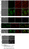
Figure 1.
Figure 1.. Imaging platelet autophagy using light microscopy.
(A) Confocal and DIC images of GFP-LC3 (GFP channel) in WT and GFP-LC3/+ mouse platelets stained live with 50 nM LysoTracker® Red DND-99 (Thermo Fisher Scientific, L7528) (Cy3 channel) at 37°C for 30 min. Images were collected on a Nikon A1R inverted confocal microscope. Arrows point to two puncta positive for both GFP-LC3 and LysoTracker. Scale bars: 5 μm. (B) Confocal and DIC images of fixed WT and GFP-LC3/+ mouse platelets incubated with PI3P-binding GST-2×FYVE and then immunostained with GST antibody (Santa Cruz, sc-459). These images show punctate structures for both GFP-LC3 and PI3P, and colocalization of GFP-LC3 and PI3P on some (labelled by arrows), but not all puncta. Recombinantly purified GST-2×FYVE was incubated at 1 μg/mL in the presence of 10 μM ZnCl2 at 4°C overnight. Controls include WT (for GFP channel) immunostained with anti-rabbit Alexa Fluor® 555 (Thermo Fisher Scientific, A31572) or anti-mouse Alexa Fluor® 647 (Thermo Fisher Scientific, A21235), but without primary antibody. Images were collected on a Nikon A1R inverted confocal microscope. Images were acquired and processed using the same settings for GFP-LC3 and WT samples. Note that in the absence of primary antibody, immunostaining with anti-mouse Alexa Fluor® 647, but not with anti-rabbit Alexa Fluor® 555, shows punctate structures in WT mouse platelets. This non-specific anti-mouse Alexa Fluor® 647-staining is likely due to non-specific binding of the anti-mouse secondary antibody to endogenous mouse immunoglobulins that are naturally packaged in the platelet α-granules. Scale bars: 5 μm. (C) Confocal and DIC images of WT and GFP-LC3/+ mouse platelets immunostained with rabbit anti-vWF (Dako, A0082) and rat anti-PF4 (R&D Systems, DY595). Note that PF4 and vWF, both of which are α-granule cargos, appear not perfectly colocalized, suggesting presence of sub-domains within the α-granules. Images were collected on a Nikon A1R inverted confocal microscope. Scale bar: 5 μm. (D) Autophagic flux in mouse platelets as monitored by GFP-LC3 puncta in the presence and absence of NH4Cl (20 mM, 2 h) using an upright wide-field microscope. Samples were visualized for DIC and GFP fluorescence (GFP-LC3). Images were acquired using a Nikon Eclipse E600 microscope equipped with a 100×/1.40 numeric aperture DIC H oil objective lens and a Zeiss AxioCam MR camera. Ten images (17 and 74 platelets/field for no treatment versus NH4Cl treatment) were obtained at random and quantified for each condition. Platelets containing GFP-LC3-positive puncta are 21 ± 8% and 64 ± 10% without and with NH4Cl treatment, respectively. Statistical significance was evaluated with the Student’s t test. This panel is reproduced from Fig. 2C in [9] with publisher’s permission.
Figure 2.
Figure 2.. Electron micrographs of autophagosome-related structures in mouse platelets.
Double-membraned phagophore-like structures (arrow heads) wrapping bulk of cytosol and/or granules in (A–B) resting and (C–D) thrombin-stimulated (0.1 U/mL, 10–30 sec) mouse platelets. Labels: m, mitochondria; α, α granules; δ, dense granules. Scale bars: 500 nm. (A–B) are reproduced from [9] with publisher’s permission.
Figure 3.
Figure 3.. Super-resolution microscopy of platelet autophagy.
3D-Structured Illumination Microscopy (SIM) images of GFP-LC3 (GFP channel) and live-stained LysoTracker Blue (405 channel, pseudo-color in red) in GFP-LC3/+ mouse platelets show incomplete ring-like GFP-LC3 structures resembling isolation membranes/phagophores as observed by electron microscopy in Fig. 2. These 3D-SIM images show an interesting spatial relationship between the GFP-LC3-positive and lysoTracker-positive structures in platelets: The lysoTracker-positive structures appear to be associated with the incomplete, ring-shaped GFP-LC3-positive structures, i.e., the lysoTracker-stained, tubular structures are either on the outside (A) or inside (B–C) of the GFP-LC3-positive rings. Images were collected on a Nikon Ti-E N-STORM/N-SIM super-resolution microscope that was equipped with an Apo SR 100X/1.49 NA oil-objective and an A1R camera. Images were processed using Nikon NIS-Elements v3.2 N-SIM/STORM software and Adobe Photoshop. Scale bars: 1 μm.

References

    1. Pease DC, An electron microscopic study of red bone marrow. Blood, 1956. 11(6): p. 501–26. - PubMed
    1. Junt T, et al., Dynamic visualization of thrombopoiesis within bone marrow. Science, 2007. 317(5845): p. 1767–70. - PubMed
    1. Machlus KR and Italiano JE Jr., The incredible journey: From megakaryocyte development to platelet formation. J Cell Biol, 2013. 201(6): p. 785–96. - PMC - PubMed
    1. Harker LA, The kinetics of platelet production and destruction in man. Clin Haematol, 1977. 6(3): p. 671–93. - PubMed
    1. Ault KA and Knowles C, In vivo biotinylation demonstrates that reticulated platelets are the youngest platelets in circulation. Exp Hematol, 1995. 23(9): p. 996–1001. - PubMed

Publication types

MeSH terms

Substances

LinkOut - more resources