

Plasma neuronal exosomes serve as biomarkers of cognitive impairment in HIV infection and Alzheimer's disease
- PMID: 30610738
- PMCID: PMC7372698
- DOI: 10.1007/s13365-018-0695-4
Plasma neuronal exosomes serve as biomarkers of cognitive impairment in HIV infection and Alzheimer's disease
Abstract
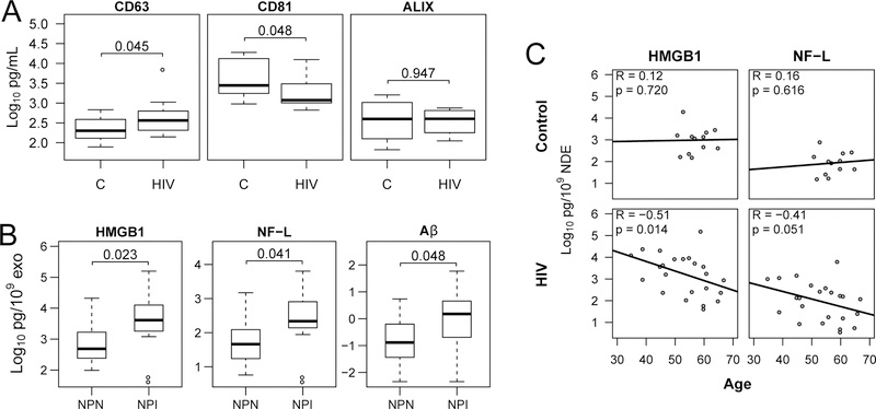
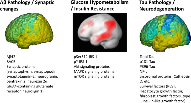
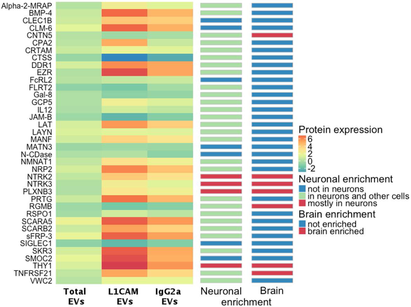
Fluid biomarkers for cognitive impairment have the advantage of being relatively noninvasive and capable of monitoring neuronal and other brain cell health in real time. Biomarkers can predict the onset of dementing illness, but also correlate with cognition in a dynamic way allowing us to follow treatment responses and determine brain recovery. Chronic HIV infection causes cognitive impairment in a subset of individuals suggesting "premature aging." Exosomes are small extracellular vesicles that are shed from all cells. They are important in normal cell-to-cell communication as they contain cellular proteins, mRNA transcripts, and miRNAs. Exosome cargo varies depending on the health of the cell and pathological state; specific proteins/mRNAs and/or miRNAs are present and may serve as biomarkers. Exosomes of variable cellular origin can be isolated from peripheral blood by various methods. Neuron-derived exosomes (NDEs) can be isolated using a precipitation/immunoaffinity approach using antibodies against neuronal cell adhesion molecule L1CAM and the contents queried for central nervous system (CNS) disorders including HIV-associated neurological disorders (HAND) and Alzheimer's disease (AD). As these studies are recent, numerous questions arise including which neuronal proteins are in NDEs and whether their contents differ in different CNS pathologies or with age. In addition, can the NDE cargo predict as well as diagnose cognitive impairment and could exosomal contents be used as therapeutic biomarkers, or theramarkers, of neuronal recovery from effective treatment? This mini-review will show some new data and review recent studies on NDE from individuals with HIV infection and AD. HIV-associated neurocognitive disorders (HAND) are pathologies seen in a subset of individuals with chronic HIV infection. They belong to the spectrum of neurodegenerative diseases that result in death or dysfunction of neurons with similarities to Alzheimer disease (AD) but also distinctive differences (reviewed (Canet et al., Front Cell Neurosci 12: 307, 2018)). Both disorders are difficult to diagnose without neuropsychological testing and both need new biomarkers to judge progression as well as recovery with treatment. Both disorders involve neuroinflammation and several common targets. AD is associated with aging and HIV is thought to initiate premature aging. In HIV infection, amyloid beta (Aβ), which is deposited in "plaques" in AD, is soluble and its relevance to HIV-associated cognitive impairment is controversial (Achim et al., J Neuroimmune Pharmacol 4: 190-199, 2009; Rempel and Pulliam, AIDS 19: 127-135, 2005). Aβ deposition is required for AD pathological diagnosis, but is not necessarily causative (Barage and Sonawane, Neuropeptides 52: 1-18, 2015; Hardy and Selkoe, Science 297: 353-356, 2002; Morris et al., Acta Neuropathol Commun 2: 135, 2014). Neurofilament light (NF-L) is a surrogate marker in plasma and cerebrospinal fluid (CSF) for neurodegeneration (Abu-Rumeileh et al., Alzheimers Res Ther 10: 3, 2018; Mattsson et al., JAMA Neurol 74: 557-566, 2017) but continues to be a controversial biomarker for both HAND and AD (Gisslen et al., EBioMedicine 3: 135-140, 2016; Kovacs et al., Eur J Neurol 24:1326-e77, 2017; Norgren et al., Brain Res 987: 25-31, 2003; Rolstad et al., J Alzheimers Dis 45: 873-881, 2015; Yilmaz et al., Expert Rev Mol Diagn 17: 761-770, 2017). Blood biomarkers are needed to advance both HAND and AD fields, as blood draws are less costly than neuroimaging and are minimally invasive compared to lumbar punctures required for CSF acquisition. Extracellular vesicles (EVs) are nanoscale membranous vesicles shed from all cells including those of the central nervous system (CNS) and found in all biofluids; they are divided into exosomes (30-150 nm) originating from late endosomes/multivesicular bodies and microvesicles (150-1000 nm) produced through budding of the plasma membrane. Both types of vesicles are implicated in the pathogenesis of neurodegenerative diseases and may provide biomarkers (Bellingham et al., Front Physiol 3: 124, 2012). In this report, we call the vesicles exosomes, since they are the predominant vesicles in our preparations. They are involved in cell-to-cell communication in normal homeostasis and can be carriers of toxic proteins (Aβ, tau) (Sardar Sinha et al., Acta Neuropathol 136: 41-56, 2018) shed by cells as waste or actively secreted in a degenerative process (review Gupta and Pulliam, J Neuroinflammation 11: 68, 2014). The idea that exosomes originating from a specific cell can be recovered in the plasma using cellular surface markers of interest is intriguing. Neuron derived exosomes (NDEs) were first described in 2015 and isolated using antibodies against neural cell adhesion molecules NCAM or L1CAM, after total plasma exosome isolation (Fiandaca et al., Alzheimers Dement 11: 600-607 e1, 2015). Characterization of NDEs follows guidelines endorsed by the International Society for Extracellular Vesicles and includes Nanoparticle Tracking Analysis (NTA) to determine EV concentration and average diameter; Western Blots for EV markers; ELISAs for neuronal proteins and transmission EM for visualization (Sun et al., AIDS 31: F9-F17, 2017; Tang et al., FASEB J 30: 3097-106, 2016). This innovative isolation of an exosome sub-population has generated interest in using NDE as biomarkers for neurodegenerative diseases like AD, HAND, traumatic brain injury, posttraumatic stress disorder and more (reviews Agoston et al., Brain Inj 31: 1195-1203, 2017; Gupta and Pulliam, J Neuroinflammation 11: 68, 2014; Hu et al., Cell Death Dis 7: e2481, 2016; Karnati et al., J Neurotrauma, 2018; Osier et al., Mol Neurobiol, 2018). Several biomarkers from plasma NDEs were recently reported by the Pulliam lab to be elevated in general cognitive impairment (Sun et al., AIDS 31: F9-F17, 2017). We review our collective data here on HAND and AD and add to the characterization of plasma NDEs as exciting biomarkers of neurodegeneration.
Keywords: Alzheimer’s; Biomarker; Dementia; Exosomes; HIV; Neurons.
Conflict of interest statement
Conflict of Interest
The authors declare that there is no conflict of interests.
Figures
References
-
- Agoston DV, Shutes-David A, Peskind ER (2017). Biofluid biomarkers of traumatic brain injury. Brain Inj 31: 1195–1203. - PubMed
-
- Anderson AM, Easley KA, Kasher N, Franklin D, Heaton RK, Zetterberg H, Blennow K, Gisslen M, Letendre SL (2018). Neurofilament light chain in blood is negatively associated with neuropsychological performance in HIV-infected adults and declines with initiation of antiretroviral therapy. J Neurovirol. - PMC - PubMed
Publication types
MeSH terms
Substances
Grants and funding
LinkOut - more resources
Full Text Sources
Other Literature Sources
Medical
Research Materials
Miscellaneous
