. 2019 Mar 1:86:312-322.
doi: 10.1016/j.actbio.2018.12.052. Epub 2019 Jan 2.

Aligned hydrogel tubes guide regeneration following spinal cord injury

Affiliations

Aligned hydrogel tubes guide regeneration following spinal cord injury

Courtney M Dumont et al. Acta Biomater. .

Abstract

Directing the organization of cells into a tissue with defined architectures is one use of biomaterials for regenerative medicine. To this end, hydrogels are widely investigated as they have mechanical properties similar to native soft tissues and can be formed in situ to conform to a defect. Herein, we describe the development of porous hydrogel tubes fabricated through a two-step polymerization process with an intermediate microsphere phase that provides macroscale porosity (66.5%) for cell infiltration. These tubes were investigated in a spinal cord injury model, with the tubes assembled to conform to the injury and to provide an orientation that guides axons through the injury. Implanted tubes had good apposition and were integrated with the host tissue due to cell infiltration, with a transient increase in immune cell infiltration at 1 week that resolved by 2 weeks post injury compared to a gelfoam control. The glial scar was significantly reduced relative to control, which enabled robust axon growth along the inner and outer surface of the tubes. Axon density within the hydrogel tubes (1744 axons/mm2) was significantly increased more than 3-fold compared to the control (456 axons/mm2), with approximately 30% of axons within the tube myelinated. Furthermore, implantation of hydrogel tubes enhanced functional recovery relative to control. This modular assembly of porous tubes to fill a defect and directionally orient tissue growth could be extended beyond spinal cord injury to other tissues, such as vascular or musculoskeletal tissue. STATEMENT OF SIGNIFICANCE: Tissue engineering approaches that mimic the native architecture of healthy tissue are needed following injury. Traditionally, pre-molded scaffolds have been implemented but require a priori knowledge of wound geometries. Conversely, hydrogels can conform to any injury, but do not guide bi-directional regeneration. In this work, we investigate the feasibility of a system of modular hydrogel tubes to promote bi-directional regeneration after spinal cord injury. This system allows for tubes to be cut to size during surgery and implanted one-by-one to fill any injury, while providing bi-directional guidance. Moreover, this system of tubes can be broadly applied to tissue engineering approaches that require a modular guidance system, such as repair to vascular or musculoskeletal tissues.

Keywords: Axon elongation; Modular biomaterial; Spinal cord injury; Tissue repair.


Conflict of interest statement

Disclosure of Potential Conflicts of Interest

The authors have no competing interests to declare.

Figures

Figure 1.
Figure 1.. Two-step polymerization can be used to generate porous macrostructures.
PEG-MAL microspheres are created with a water/oil emulsion method using a plasmin sensitive YKND peptide crosslinker. Scale bar 100 μm. (A). (B) Resulting microspheres had a diameter distribution ranging from 15-105 μm with an average size of 45 μm. (C) Tubes and bridges can be fabricated from the microspheres using UV-sensitive I2959 photoinitiator to crosslink remaining open MAL side arms. Tubes can be subsequently formed into a bridge composite using fibrin hydrogel to hold the tubes together during implantation.
Figure 2.
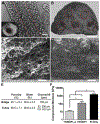
Figure 2.. Tertiary structures, such as tubes and bridges can be created using a second crosslinking phase that capitalizes on MAL sidechains that were not utilized during microsphere fabrication.
Resulting tubes (A – longitudinal, inset - transverse, scale bars 100 μm) and bridges (B, scale bar 200 μm) generated from the PEG-MAL microspheres are porous and contain aligned channels within the material. Using SEM, pores can be seen through the hydrogel tubes (C, 50 μm scale) and bridges (D, 50 μm scale), albeit the pore structure does appear to vary between the two structures. (E) No significant differences in the porosity, swelling ratio, or water retention capacity were detected between the two structures. (F) Young’s modulus for PEG-MAL crosslinked with YKND/PI using microspheres in a two-step polymerization technique (12.52 kPa), as used to generate tubes and bridges, was significantly lower (p < 0.0001) than the modulus for PEG-MAL crosslinked with YKND/PI without first forming microspheres (129.2 kPa) and PI only (1588 kPa).
Figure 3.
Figure 3.. Immune cell infiltration 7 days after injury.
Gelfoam alone (negative control) or with a biomaterial implant (PEG bridges or PEG tubes) was implanted into a lateral hemisection spinal cord injury (A) and apposition to intact tissue was visually verified (B). GFAP+ astrocytes (C) and CD45+ leukocytes (D) infiltrate the injury site, however, no difference in the bulk populations of these cells are evident across the blank (gelfoam) injury or PEG scaffolds. (E) Further evaluation of the leukocyte phenotypes including CD11c+ DCs, F4/80+ macrophages, F4/80+arginase+ M2 macrophages (teal overlay), Lyg6+ neutrophils, and CD4+ T-cells identified a significant increase in DCs and macrophages within the PEG materials. Data are represented as mean ± standard deviation. n = 5, * p < 0.05.
Figure 4.
Figure 4.. Glial scar thickness 2 weeks after injury.
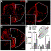
GFAP+ astrocytes were observed throughout the intact tissue with robust staining at the interface of the gelfoam (A), bridge (B), and tube (C) implants with the intact tissue. The rostral margin thickness (bottom image outlined in red for panels A-C) was measured at multiple locations for each tissue. (D) PEG implants significantly reduced glial scar thickness (p < 0.01) compared to the gelfoam control. Data are represented as mean ± standard deviation. n= 3, ** p < 0.01. 500 μm, 100 μm (inset) scale bars.
Figure 5.
Figure 5.. PEG implants enhance axon elongation at 8 weeks after transplantation in T9-10 hemisection
Qualitatively, NF-200+ (red) axon expression is greater in PEG bridge (B) and tube (C) implants compared to gelfoam (A). (D) Quantification of regenerating axons was binned into three 0.75 mm lateral sections of the implant: rostral (R), middle of the injury (M), and caudal (C) as depicted in the schematic inset. Axon density is increased at both the rostral and caudal segments in mice receiving 5-tube composites, while the axon density for mice receiving the hydrogel bridges only increased at the rostral margin compared to gelfoam at 8 weeks post injury. Data are represented as mean ± standard deviation . n = 6, ** p < 0.01. 200 μm scale bar.
Figure 6.
Figure 6.. Axon myelination is supported in all conditions at 8 weeks post injury.
(A) NF-200+ axons co-localized with oligodendrocyte derived myelin (MBP+; green) and with Schwann cell myelin (MBP+P0+; blue) within transverse sections of the bridge across all conditions. Unmyelinated axons (denoted by *), oligodendrocyte myelinated axons (denoted by ^), and Schwann cell myelinated axons (denoted by ▴) are observed in each condition. Nodes between myelinated areas of axons can also be observed (denoted by arrows). No significant difference was observed in the density of myelinated axons (B), however, a significant decrease in percentage of myelinated axons was observed in the bridge and tube conditions compared to gelfoam (C). (D) While the overall percent of myelination was lower, both PEG tubes and bridges did result in a significant increase in the percentage of oligodendrocyte-derived myelin (NF-200+MBP+P0; p < 0.05) compared to gelfoam. Data are represented as mean ± standard deviation . n = 6, * p < 0.05. 10 μm scale bar.
Figure 7.
Figure 7.. Improved hindlimb locomotor abilities achieved with PEG implants.
An open field locomotor test known as the BMS was used to asses mouse hindlimb mobility for 8 weeks after injury and biomaterial implantation. By post-operative week 2, mice with PEG bridges achieved a significantly higher average BMS score compared to mice with gelfoam (* significance of bridges compared to gelfoam; p < 0.05, n = 6). Mice with PEG tubes exhibited a significant increase in BMS score by week 4 compared to gelfoam (^ significance of bridges compared to gelfoam; p < 0.05, n = 6).

References

    1. Xu T, Zhao W, Zhu JM, Albanna MZ, Yoo JJ, Atala A, Complex heterogeneous tissue constructs containing multiple cell types prepared by inkjet printing technology, Biomaterials 34(1) (2013) 130–9. - PubMed
    1. Mapili G, Lu Y, Chen S, Roy K, Laser-layered microfabrication of spatially patterned functionalized tissue-engineering scaffolds, J Biomed Mater Res B Appl Biomater 75(2) (2005) 414–24. - PubMed
    1. Raineteau O, Schwab ME, Plasticity of motor systems after incomplete spinal cord injury, Nat Rev Neurosci 2(4) (2001) 263–73. - PubMed
    1. Anderson KD, Cowan RE, Horsewell J, Facilitators and Barriers to Spinal Cord Injury Clinical Trial Participation: Multi-National Perspective of People Living with Spinal Cord Injury, J Neurotrauma 33(5) (2016) 493–9. - PubMed
    1. Basso DM, Beattie MS, Bresnahan JC, Graded histological and locomotor outcomes after spinal cord contusion using the NYU weight-drop device versus transection, Exp Neurol 139(2) (1996) 244–56. - PubMed

Publication types

MeSH terms