

Irreversible electroporation in a case of pancreatic leiomyosarcoma: a novel weapon versus a rare malignancy?
- PMID: 30611280
- PMCID: PMC6320590
- DOI: 10.1186/s12957-018-1553-9
Irreversible electroporation in a case of pancreatic leiomyosarcoma: a novel weapon versus a rare malignancy?
Abstract
Background: Primary pancreatic leiomyosarcoma is an extremely rare entity that needs high clinical suspicion in order to diagnose it at an early stage. Clinical characteristics, diagnosis, and management still remain challenging and controversial, especially in advanced stages, when tumor invades adjacent vessels and organs or gives distant metastases.
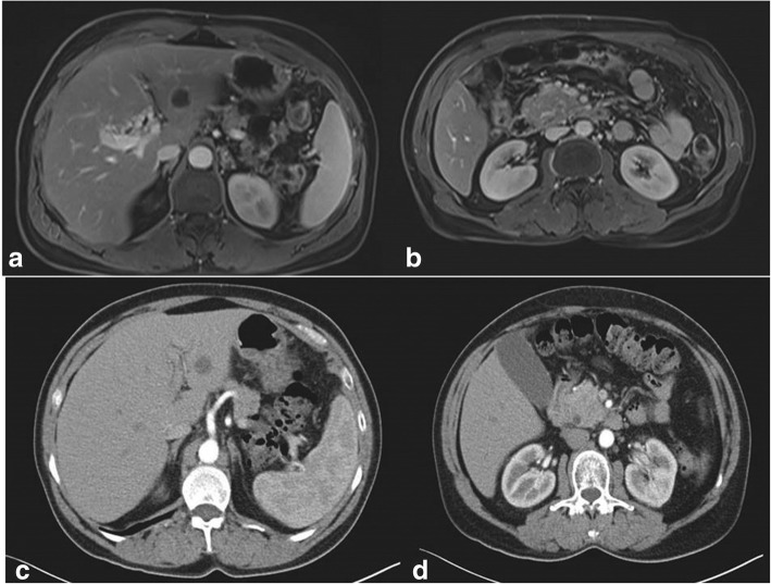
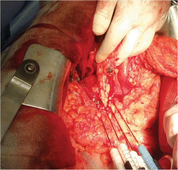
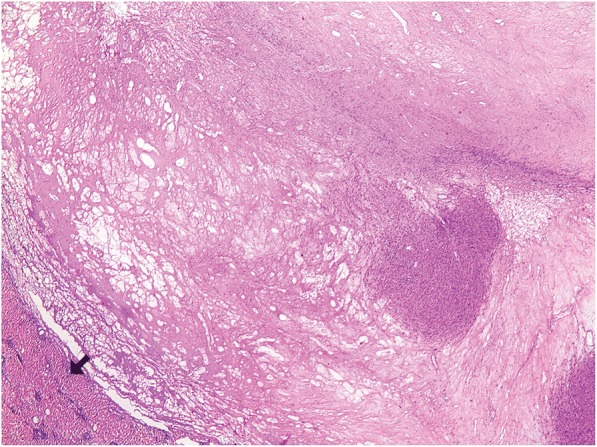
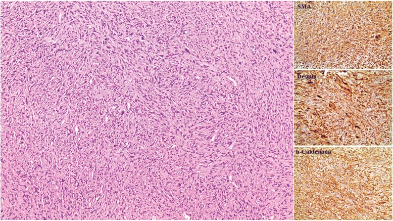
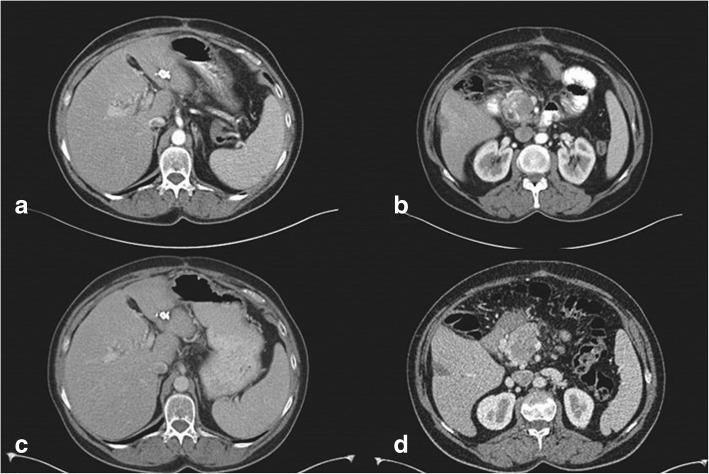
Case presentation: Herein, we describe a case of a 57-year-old woman suffering from advanced pancreatic leiomyosarcoma with thrombosis of the superior mesenteric vein, as well as liver lesions which were suspicious for metastasis. Multidisciplinary team decided for upfront chemotherapy to assess tumor response. Follow-up imaging after the completion of chemotherapy led tumor board to decide for subsequent surgical exploration. The patient underwent exploratory laparotomy and irreversible electroporation ablation of the pancreatic tumor. Postoperative course was uneventful, and she was discharged 10 days later with a plan to receive adjuvant therapy. To the best of our knowledge, this is the first case of pancreatic leiomyosarcoma ever reported, treated with this novel technique of irreversible electroporation that could be an alternative and feasible way for the management of these rare malignancies.
Conclusions: In conclusion, primary pancreatic leiomyosarcoma is a rare and highly malignant tumor associated with poor prognosis. Nowadays, R0 surgical resection remains the cornerstone treatment, combined with adjuvant and/or neoadjuvant chemotherapy prior to resection. In the advanced setting, when major vessel invasion and distant metastases occur, chemotherapy along with irreversible electroporation ablation could be a helpful and possibly effective modality for the management of this highly aggressive tumor.
Keywords: IRE; Irreversible electroporation; Pancreatic cancer; Pancreatic leiomyosarcoma.
Conflict of interest statement
Ethics approval and consent to participate
Not applicable
Consent for publication
Informed consent was obtained from the patient.
Competing interests
The authors declare that they have no competing interests.
Publisher’s Note
Springer Nature remains neutral with regard to jurisdictional claims in published maps and institutional affiliations.
Figures
References
Publication types
MeSH terms
LinkOut - more resources
Full Text Sources
Medical
