. 2019 Mar 21;26(3):390-399.e5.
doi: 10.1016/j.chembiol.2018.11.005. Epub 2019 Jan 3.

What Makes a Kinase Promiscuous for Inhibitors?

Affiliations

What Makes a Kinase Promiscuous for Inhibitors?

Sonya M Hanson et al. Cell Chem Biol. .

Abstract

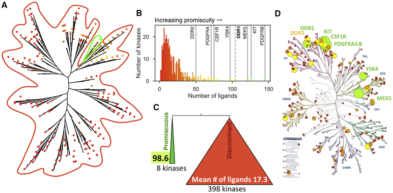
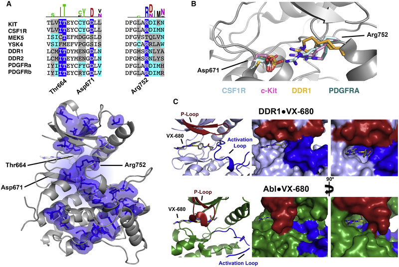
ATP-competitive kinase inhibitors often bind several kinases due to the high conservation of the ATP binding pocket. Through clustering analysis of a large kinome profiling dataset, we found a cluster of eight promiscuous kinases that on average bind more than five times more kinase inhibitors than the other 398 kinases in the dataset. To understand the structural basis of promiscuous inhibitor binding, we determined the co-crystal structure of the receptor tyrosine kinase DDR1 with the type I inhibitors dasatinib and VX-680. Surprisingly, we find that DDR1 binds these type I inhibitors in an inactive conformation typically reserved for type II inhibitors. Our computational and biochemical studies show that DDR1 is unusually stable in this inactive conformation, giving a mechanistic explanation for inhibitor promiscuity. This phenotypic clustering analysis provides a strategy to obtain functional insights not available by sequence comparison alone.

Keywords: ABL; DDR1; DFG; Folding@home; Markov state model; crystallography; drug promiscuity; kinase inhibition; molecular dynamics; selectivity.


Conflict of interest statement

Declaration of interests

J.D.C. is a member of the S.A.B. of Schrödinger LLC.

Figures

Figure 1:
Figure 1:. Hierarchical clustering of the human kinome by inhibition phenotype reveals a group of highly promiscuous kinases.
A) Hierarchical clustering analysis of the PKIS2 dataset assessing KinoBead binding inhibition for 406 kinases by 645 ligands at 1 μM concentration. Individual kinases are shown as dots and colored by the number of inhibitors capable of displacing more than 90% of kinase from covalently-tethered pan-kinase inhibitors (green being most inhibitors, red being fewest inhibitors). The promiscuous branch is circled in green. B) The most basal branch of this dendrogram is a single branch of eight promiscuous kinases that bind a mean number of 98.6 ± 31.9 ligands (green) while the other 398 in the panel bind 17.3 ± 14.0 ligands (red) (mean ± SD). C) Number of kinases that are inhibited by given number of inhibitors to more than 90%. D) Superposition of the promiscuity of kinases onto the kinase phylogenetic tree. Circle diameter is proportional to kinase promiscuity for emphasis, and colors are as in C. See also Figures S1 and S2.
Figure 2:
Figure 2:. DDR1 binds type I inhibitors in the DFG-Asp-out conformation.
A) Co-crystal structure of DDR1·VX-680 [PDB-entry: 6BRJ] with activation loop (blue), phosphate binding P-loop (red), helix αC (orange). B) The co-crystal structure of DDR1·dasatinib [PDB-entry: 6BSD] with regulatory elements colored as in A. C) Comparison of type I (top, VX-680/dasatinib) and type II (bottom, imatinib/ponatinib) inhibitors binding to Abl (green, right column) and DDR1 kinase (blue, left column). Only the protein surrounding the DFG-motif is shown for clarity. D) Comparison of Φ dihedral angle for Asp747 in DDR1 (blue) and Abl (green) bound to the type I and type II inhibitors in panel C. See also Figure S3.
Figure 3:
Figure 3:. Wild-type DDR1 is stable in the DFG-Asp-out conformation and mutations destabilize this inactive conformation.
A) Snapshots from one of the trajectories seen to flip from DFG-Asp-out to DFG-Asp-in are superimposed on the final free energy landscape for WT DDR1 are highlighted in yellow. HMM macrostates representing DFG-in (red) and DFG-out (purple) states are shown, and transparency is proportional to the membership of each k-means cluster center to the macrostate. B) The Asp671-Arg752 salt bridge, Asp671-Tyr755 hydrogen bond, and Asp729-Tyr759 interaction thought to stabilize the inactive DFG-Asp-out conformation. C) Free energy landscapes for WT, D671N, Y755A, and Y759A simulations superimposed onto the first two (slowest) TICs of the WT simulations. Dotted line corresponds to the linear separation between states seen in C. D) Free energy difference between DFG-out and DFG-in states calculated from the free energy landscapes in panel C. Positive values indicate stabilization in the DFG-out conformation; negative values indicate stabilization in the DFG-in conformation. Error bars indicate 95% confidence intervals from a Bayesian MSM with 1000 samples. E) Kinase activity assays of DDR1. F) Binding affinity of DDR1 wt and mutant proteins for the type II inhibitor imatinib. Binding affinity for imatinib is reported as inhibitory constant Ki for competition with a general kinase inhibitor. See also Figure S4 and Table S1.
Figure 4:
Figure 4:. Structural hallmarks of DDR1 and other promiscuous kinases.
A) Sequence alignment of the eight promiscuous kinases around Thr664 (the ‘gatekeeper’ mutant), Asp671, and Arg752 (DDR1 numbering). Dark blue highlights residues enriched in the promiscuous set by 50% or more and cyan indicates residues enriched by 10–50% according to Two Sequence Logo comparison (using p-value cutoff 0.01) of the promiscuous kinases to the other 483 kinases analyzed. Bottom: All residues enriched in our promiscuous set by 50% or more are shown as blue surface on the DDR1-VX680 structure. B) Potential salt bridges between residues equivalent to DDR1 Asp671 and Arg752 are shown for representative structures: CSF1R (PDB IDs: 3KRL and 2IOV) in pale cyan, c-Kit (PDB IDs: IT45 and IT46) in pink, DDR1 (PDB IDs: 5FDP and 5BVO) in orange, and PDGFRA (PDB ID: 5GRN) in teal. C) The structures of VX-680 bound to DDR1 (top) and Abl (bottom, PDB entry 2F4J) illustrate the hydrophobic shield formed by the activation loop (blue) and the P-loop (red), respectively. See also Figure S5.

References

    1. Adrián FJ, Ding Q, Sim T, Velentza A, Sloan C, Liu Y, Zhang G, Hur W, Ding S, Manley P, et al. (2006). Allosteric inhibitors of Bcr-abl-dependent cell proliferation. Nature Chemical Biology 2, 95–102. - PubMed
    1. Anastassiadis T, Deacon SW, Devarajan K, Ma H, and Peterson JR (2011). Comprehensive assay of kinase catalytic activity reveals features of kinase inhibitor selectivity. Nature Biotechnology 29, 1039–1045. - PMC - PubMed
    1. Artim SC, Mendrola JM, and Lemmon MA (2012). Assessing the range of kinase autoinhibition mechanisms in the insulin receptor family. The Biochemical journal 448, 213–220. - PMC - PubMed
    1. Azam M, Latek RR, and Daley GQ (2003). Mechanisms of autoinhibition and STI-571/imatinib resistance revealed by mutagenesis of BCR-ABL. Cell 112, 831–843. - PubMed
    1. Canning P, Tan L, Chu K, Lee SW, Gray NS, and Bullock AN (2014). Structural Mechanisms Determining Inhibition of the Collagen Receptor DDR1 by Selective and Multi-Targeted Type II Kinase Inhibitors. Journal of molecular biology. - PMC - PubMed

Publication types

MeSH terms