. 2018 Nov 13;8(21):5986-5994.
doi: 10.7150/thno.26650. eCollection 2018.

Chemoresistance Transmission via Exosome-Mediated EphA2 Transfer in Pancreatic Cancer

Affiliations

Chemoresistance Transmission via Exosome-Mediated EphA2 Transfer in Pancreatic Cancer

Jia Fan et al. Theranostics. .

Abstract

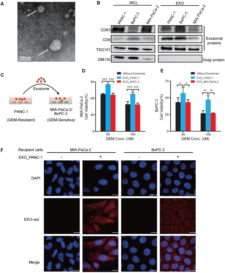
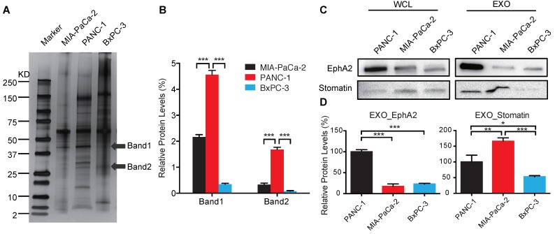
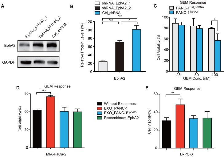
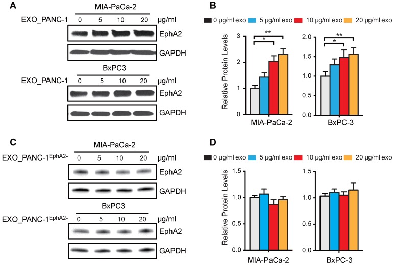
Rationale: Exosomes are small extracellular vesicles secreted by most cells that are found in blood and other bodily fluids, and which contain cytoplasmic material and membrane factors corresponding to their cell type of origin. Exosome membrane factors and contents have been reported to alter adjacent and distant cell behavior in multiple studies, but the impact of cancer-derived exosomes on chemoresistance is less clear. Methods: Exosomes isolated from three pancreatic cancer (PC) cell lines displaying variable gemcitabine (GEM) resistance (PANC-1, MIA PaCa-2, and BxPC-3) were tested for their capacity to transmit chemoresistance among these cell lines. Comparative proteomics was performed to identify key exosomal proteins that conferred chemoresistance. Cell survival was assessed in GEM responsive PC cell lines treated with recombinant Ephrin type-A receptor 2 (EphA2), a candidate chemoresistance transfer factor, or exosomes from a chemoresistant PC cell line treated with or without EphA2 shRNA. Results: Exosomes from chemoresistant PANC-1 cells increased the GEM resistance of MIA PaCa-2 and BxPC-3 cell cultures. Comparative proteomics determined that PANC-1 exosomes overexpressed Ephrin type-A receptor 2 (EphA2) versus exosomes of less chemoresistant PC cell lines MIA PaCa-2 and BxPC-3. EphA2-knockdown in PANC-1 cells inhibited their ability to transmit exosome-mediated chemoresistance to MIA PaCa-2 and BxPC-3, while treatment of MIA PaCa-2 and BxPC-3 cells with soluble EphA2 did not promote chemoresistance, indicating that membrane carried EphA2 was important for the EphA2 chemoresistance effect. Conclusion: Exosomal EphA2 expression could transmit chemoresistance and may potentially serve as a minimally-invasive predictive biomarker for PC treatment response. Further work should address whether additional exosomal factors regulate resistance to other cancer therapeutic agents for PC or other cancer types.

Keywords: Cytotoxic resistance; EphA2; Exosome; Gemcitabine; Pancreatic cancer.


Conflict of interest statement

Competing Interests: The authors have declared that no competing interest exists.

Figures

Figure 1
Figure 1
Exosomes of GEM-resistant PC cells can transfer chemoresistance. (A) TEM image of exosomes isolated from PANC-1 cells and negatively stained by uranyl acetate (arrows). (B) Western blot analysis of exosomes (EXOs) and whole-cell lysates (WCLs) of PANC-1, MIA PaCa-2, and BxPC-3 cells for exosome (CD63, CD9, and TSG101) and golgi (GM130) protein markers. (C) Experimental design of exosome uptake studies. (D-E) GEM cytotoxicity in (D) MIA PaCa-2 cells or (E) BxPC-3 after 24 h pre-treatment with 20 µg/mL exosomes from PANC-1 (EXO_PANC-1), BxPC-3 (EXO_BxPC-3) or MIA PaCa-2 (EXO_MIA PaCa-2) cells, with fresh exosomes added every 24 h of the 3 day GEM treatment. Cell viability is presented as a percentage of control (no drug or exosomes) viability. (F) Exosome internalization in MIA PaCa-2 and BxPC-3 cells incubated for 2 h with or without EXO-Red-stained exosomes and then stained with DAPI for nuclear visualization. Bar indicates 10 µm; Data indicate mean±SD; n=6; *p<0.05; **p<0.01, ***p<0.001.
Figure 2
Figure 2
Identification of candidate exosome chemoresistance proteins. (A) Western blot of exosomal proteins of MIA PaCa-2, PANC-1, and BxPC-3 cells (arrows indicate protein bands subjected to LC-MS/MS sequence analysis). (B) Relative expression of selected exosomal protein bands normalized to the sum of the band densities in each lane. (C) Western blot of candidate proteins in WCLs and EXOs of PANC-1, MIA PaCa-2, and BxPC-3 cells. (D) Relative exosome EphA2 and Stomatin expression. Data indicate mean±SD (n=3); *p<0.05, **p<0.01, ***p<0.001.
Figure 3
Figure 3
Exosomal EphA2 confers GEM resistance. (A) Western blot and (B) GAPDH-normalized EphA2 levels in PANC-1 cells transduced with lentiviruses expressing EphA2_shRNA_1, EphA2_shRNA_2, or Ctrl_shRNA. (C) Viability of PANC-1Ctrl_shRNA and PANC-1EphA2- (EphA2_shRNA_1 transduced) cells after 72h GEM treatment, normalized to untreated cell viability. (D-E) GEM cytotoxicity (100nM for 72 h) in (D) MIA PaCa-2 and (E) BxPC-3 cells after exposure to 20 µg/mL EXO_PANC-1 or EXO_PANC-1EphaA2- or recombinant EphA2 (0.5 µg/mL). Data represent mean±SD; Western: n=3; MTT assay: n=6; *p<0.05, **p<0.01, ***p<0.001.
Figure 4
Figure 4
Exosomal EphA2 expression does not affect exosome internalization. (A) Confocal images of MIA PaCa-2 and BxPC-3 cells mock-treated or incubated for 2 h with the indicated EXO-Red-stained exosomes, then fixed, permeabilized, and stained with DAPI (blue). The scale bar represents 10 µm. (B-C) Flow cytometric analysis of the fluorescence intensity of (B) BxPC-3 and (C) MIA PaCa-2 cells after 2h co-culture with the indicated EXO-Red-labeled exosomes.
Figure 5
Figure 5
EphA2 uptake by recipient PC cells. (A and C) Western blots of MIA PaCa-2 and BxPC3 WCLs after 96 h culture with (A) PANC-1 or (C) PANC-1EphA2- exosomes. (B and D) GAPDH-normalized EphA2 expression. Data represent mean±SD; n=3; *p<0.05, **p<0.01, ***p<0.001.

References

    1. Tredan O, Galmarini CM, Patel K, Tannock IF. Drug resistance and the solid tumor microenvironment. J Natl Cancer Inst. 2007;99:1441–54. - PubMed
    1. Swartz MA, Iida N, Roberts EW, Sangaletti S, Wong MH, Yull FE. et al. Tumor microenvironment complexity: emerging roles in cancer therapy. Cancer Res. 2012;72:2473–80. - PMC - PubMed
    1. Vincent A, Herman J, Schulick R, Hruban RH, Goggins M. Pancreatic cancer. Lancet. 2011;378:607–20. - PMC - PubMed
    1. Burris HA 3rd, Moore MJ, Andersen J, Green MR, Rothenberg ML, Modiano MR. et al. Improvements in survival and clinical benefit with gemcitabine as first-line therapy for patients with advanced pancreas cancer: a randomized trial. J Clin Oncol. 1997;15:2403–13. - PubMed
    1. Ryan DP. Chemotherapy for advanced exocrine pancreatic cancer. UpToDate; 2015.

Publication types