Review
. 2018 Nov 29;8(22):6210-6232.
doi: 10.7150/thno.26610. eCollection 2018.

PET-MR and SPECT-MR multimodality probes: Development and challenges

Affiliations
Review

PET-MR and SPECT-MR multimodality probes: Development and challenges

Chang-Tong Yang et al. Theranostics. .

Abstract

Positron emission tomography (PET)-magnetic resonance (MR) or single photon emission computed tomography (SPECT)-MR hybrid imaging is being used in daily clinical practice. Due to its advantages over stand-alone PET, SPECT or MR imaging, in many areas such as oncology, the demand for hybrid imaging techniques is increasing dramatically. The use of multimodal imaging probes or biomarkers in a single molecule or particle to characterize the imaging subjects such as disease tissues certainly provides us with more accurate diagnosis and promotes therapeutic accuracy. A limited number of multimodal imaging probes are being used in preclinical and potential clinical investigations. The further development of multimodal PET-MR and SPECT-MR imaging probes includes several key elements: novel synthetic strategies, high sensitivity for accurate quantification and high anatomic resolution, favourable pharmacokinetic profile and target-specific binding of a new probe. This review thoroughly summarizes all recently available and noteworthy PET-MR and SPECT-MR multimodal imaging probes including small molecule bimodal probes, nano-sized bimodal probes, small molecular trimodal probes and nano-sized trimodal probes. To the best of our knowledge, this is the first comprehensive overview of all PET-MR and SPECT-MR multimodal probes. Since the development of multimodal PET-MR and SPECT-MR imaging probes is an emerging research field, a selection of 139 papers were recognized following the literature review. The challenges for designing multimodal probes have also been addressed in order to offer some future research directions for this novel interdisciplinary research field.

Keywords: PET-MR; SPECT-MR; bimodality imaging probe; contrast agent; radioligand.


Conflict of interest statement

Competing Interests: The authors have declared that no competing interest exists.

Figures

Figure 1
Figure 1
Bimodal probe Gd-DOTA-4AMP-F. Adapted with permission from , copyright 2010 Wiley-VCH.
Figure 2
Figure 2
Metal complexes and 68Ga labeling of TRAP(HMDA-DOTA)3 (1). Adapted with permission from , copyright 2013 Wiley-VCH.
Figure 3
Figure 3
Schematic representation of the two Gd3+ and Ho3+ complexes. Adapted with permission from , copyright 2011 Royal Society of Chemistry.
Figure 4
Figure 4
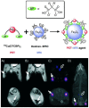
(Left) Schematic representation of dual-modality 64Cu-mSPIO. (Right) PET and MR imaging of 64Cu-mSPIO phantoms. (A) T2-weighted image of 25 µg Fe/mL (bottom) of 64Cu-labeled magnetic nanoparticles (scale bar corresponds to high and low iron concentration). (B) Decay-corrected microPET image of 25 µg Fe/mL (top) and 10 µg Fe/mL (bottom) of 64Cu-labeled magnetic nanoparticles (scale bar corresponds to high and low copper-64 concentration). Adapted with permission from , copyright 2010 American Chemical Society.
Figure 5
Figure 5
Illustration of PET-MRI probe based on IO nanoparticles. Adapted with permission from , copyright 2008 Society of NuclearMedicine and Molecular Imaging.
Figure 6
Figure 6
(Left) Decay-corrected whole-body coronal PET images of nude mice bearing human U87MG tumor at 1, 4, and 21 h after injection of 3.7 MBq of 64Cu-DOTA-IO, 64CU-DOTA-IO-RGD, or 64Cu-DOTA-IO-RGD with 10 mg of c(RGDyK) peptide per kg (300 µg of iron-equivalent IO particles per mouse). (Right) T2-weighted MR images of nude mice bearing U87MG tumor before injection of IO nanoparticles (A and E) and at 4 h after tail-vein injection of DOTA-IO (B and F), DOTA-IO-RGD (C and G), and DOTA-IO-RGD with blocking dose of c(RGDyK) (D and H). Adapted with permission from , copyright 2008 Society of Nuclear Medicine and Molecular Imaging
Figure 7
Figure 7
(a-f) PET-MR images of SLNs in a rat at 1 h post injection of 124I-SA-MnMEIO into the right forepaw (I = nanoprobe injection site). Coronal a) MR and b) PET images in which a brachial LN (white circle) is detected. c) The position of the brachial LN is well matched in a PET-MR fusion image. Four small pipette tips containing Na124I solution were used as a fiducial marker (white arrowheads) for the concordant alignment in PET-MR images. In the transverse images, axillary (red circle) and brachial LNs (white circle) are detected in the d) MR and e) PET images, and images of each node are nicely overlapped in the corresponding PET/MR fusion image (f). g) The explanted brachial LN also shows consistent results with in vivo images by PET and MR. Only the LN from the right-hand side of the rat containing 124I-SA-MnMEIO shows strong PET and dark MR images. The schematics of the rat in the h) coronal and i) transverse directions show the locations of the LNs. Adapted with permission from , copyright 2008 Wiley-VCH.
Figure 8
Figure 8
(Top) Radiolabeling of dextran-coated iron oxide nanoparticles such as Endorem using radiolabeled bisphosphonates (BPs). (Bottom) In vivo PET-MR imaging studies with [64Cu(dtcbp)2]-Endorem in a mouse. (A, B) Coronal (top) and short axis (bottom) MR images of the lower abdominal area and upper hind legs showing the popliteal lymph nodes (solid arrows) before (A) and after (B) footpad injection of [64Cu(dtcbp)2]-Endorem. (C) Coronal (top) and short-axis (bottom) NanoPET-CT images of the same mouse as in (B) showing the uptake of [64Cu(dtcbp)2]-Endorem in the popliteal (solid arrow) and iliac lymph nodes (hollow arrow). (D) Whole-body NanoPET-CT images showing sole uptake of [64Cu(dtcbp)2]-Endorem in the popliteal and iliac lymph nodes. No translocation of radioactivity to other tissues was detected. Adapted with permission from , copyright 2011 Wiley-VCH.
Figure 9
Figure 9
Long-circulating bimodal nanoparticles for PET-MR and SPECT-MRI. (A) Bisphosphonate anchors allow strong and stable binding of PEG polymers and radionuclides on the surface of the USPIOs. (B) The bimodal nanoparticles circulate in the bloodstream, as indicated by the strong imaging signal in the heart and vessels. Adapted with permission from , copyright 2013 American Chemical Society.
Figure 10
Figure 10
Schematic of the carboxyl-terminated polyglucose sorbitol carboxymethylether coating (grey) surrounding the iron oxide crystal core (green) of the nanoparticle. Amination of the particles was carried out before their functionalization with ~7 DFO chelates. Adapted with permission from , copyright 2014 Springer Nature.
Figure 11
Figure 11
Reaction of FH with 89Zr4+ ion salts (oxalate or chloride) to give radiolabeled 89Zr-FH. Adapted with permission from , copyright 2015 Royal Society of Chemistry.
Figure 12
Figure 12
(Left) A schematic illustration of chelator-free synthesis of 69Ge-metal oxides. (Rignt) a) In vivo lymph node imaging with PET after subcutaneous injection of 69Ge-SPION@PEG into the left footpad of the mouse. Lymph nodes and paws are indicated by green and red arrows, respectively. b) Quantification of the 69Ge-SPION@PEG uptake in the lymph node and mouse paw (n = 3). c) In vivo lymph node mapping with MRI before and after injection of Ge-SPION@PEG into the left foot pad of the mouse. Obvious darkening of the lymph node can be seen (dashed green circle), whereas no contrast enhancement is observed for the contralateral lymph node (dashed red circle). Adapted with permission from , copyright 2014 Wiley-VCH.
Figure 13
Figure 13
Radio-labelling of SPION-MWNT hybrids with technetium-99m (99mTc) via a linker. The Radio-labelling was conducted using 99mTc-dipicolylamine-alendronate (99mTc-DPAale). Adapted with permission from , copyright 2014Wiley-VCH.
Figure 14
Figure 14
Final steps in the synthesis of AGuIX nanoparticles: (a) nanoparticles after transfer in water; (b) addition of DTPA ligands; (c) chelation of Gd3+ previously trapped in NODAGA ligands; (d) possibility of radiolabelling with 68Ga on accessible NODAGA ligands. On the left is the dynamic light scattering distribution of AGuIX and on the right is their mass distribution determined by deconvolution with a multiplicative correlation algorithm. Adapted with permission from , copyright 2011Wiley-VCH.
Figure 15
Figure 15
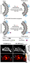
(Top) LP constructs and radiolabeling strategy: OCT was conjugated to Gd-Control LPs (CL) resulting in targeted OCT-LP (OL). Both paramagnetic formulations were radiolabeled using 89Zr with a chelator-free approach. (Bottom) PET-MR co-registration: MR (top row) and PET (bottom) scans of mice bearing wild type (wt) (→) and SSTr2-postive tumors (♦→) and injected with CL (left panel) and OL (right panel). Adapted with permission from , copyright 2013 Springer Nature.
Figure 16
Figure 16
Small molecule trimodal probe. Adapted with permission from , copyright 2011 Loyal Society of Chemistry.
Figure 17
Figure 17
Multimodality molecular imaging of melanin nanoparticles. The melanin granules were first dissolved in 0.1 N NaOH aqueous solution, and then neutralized under sonication to obtain melanin nanoparticles with high water monodispersity and homogeneity. After PEG surface-modification, RGD was further attached to the MNP for tumor targeting. Then, Fe3+ and/or 64Cu2+ were chelated to the obtained MNPs for PAI/MRI/PET multimodal imaging. Adapted with permission from , copyright 2014 American Chemical Society.
Figure 18
Figure 18
Schematic diagram of triple-labeled CLIO-Tat. The developed magnetic particle consists of a central superparamagnetic iron oxide core (yellow), sterically shielded by crosslinked dextran (green). The particle core measures ~5 nm and the overall particle size is 45 nm. The FITC-derivatized Tat peptide (blue) was attached to the aminated dextran, yielding an average four peptides per particle. The dextran surface was also modified with the chelator DTPA (red) for isotope labeling. Adapted with permission from , copyright 2000 Springer Publishing Ltd.
Figure 19
Figure 19
Triple-modality imaging of radiolabeled nanoparticles: a) optical, b) microPET, and c) MRI of 124I-labeled SPIONs injected into the front paws of a BALB/c mouse bearing a 4T1 tumor implanted on its shoulder. Tumor: yellow arrow; sentinel lymph node: red dotted circle; injection site: “I”; bladder: red arrow; fiduciary markers: white arrow head. d) Ex vivo luminescence (top) and microPET (bottom) images of the dissected lymph nodes. e) Schematic diagram of the tumor metastasis model and injection route of radiolabeled nanoparticles. Adapted with permission from , copyright 2010 Wiley-VCH.
Figure 20
Figure 20
Synthesis scheme of 124I-(cRGDyk)2-UCNP conjugates. Polymer-coated UCNP (pcUCNP) was reacted both with (cRGDyk)2 peptide and with MeO-PEGNH2 (molecular weight, 2,000) using EDC/Nhydroxysulfosuccinimide. Tyrosine residues of (cRGDyk)2 were labeled with 124I using Iodo-Beads (Pierce). Adapted with permission from , copyright 2013 Society of Nuclear Medicine and Molecular Imaging.
Figure 21
Figure 21
Lymph node PET-MRI imaging of a mouse with an inflamed right leg using 18F-labeled Fe3O4@NaYF4(Yb, Tm)-BP-PEG NPs (a-d) or with [18F]-fluoride only (e-g): (a) whole-body PET image showing uptake of radiolabeled NPs (maximum intensity projection; bone uptake was observed due to gradual release of fluoride from NPs due to the 7 h delay post injection of NPs); (b) PET image showing popliteal and iliac lymph nodes (coronal section); (c) PET-MRI fused image (coronal section); (d) MR image (coronal section) with darkening contrast inside the popliteal lymph node at left-rear (white circle) and “outside” lymph node at the inflamed right-rear (red circle) induced by injection of 30 μL 0.67 mg/mL lipopolysaccharide (LPS) 18 h prior to imaging, and at iliac lymph node; (e) PET image following injection of [18F]-fluoride showing no contrast in lymph nodes in the absence of NPs and prominent uptake by skeleton; (f) PET-MRI fused image following injection of [18F]-fluoride, showing no radioactivity associated with lymph nodes; (g) MR image showing no difference between normal popliteal lymph node at left-rear leg (white circle) and the inflamed lymph node at right-rear leg induced by injection of 30 μL 0.67 mg/mL LPS 18 h prior to imaging; and (h-k) enlarged MR images of corresponding lymph nodes. Adapted with permission from , copyright 2016 American Chemical Society.

Similar articles

Cited by

References

    1. Mankoff DA. A definition of molecular imaging. J Nucl Med. 2007;48(N):18–21. - PubMed
    1. Markus R. Molecular imaging: basic principles and applications in biomedical research: World Scientific. 2013.
    1. Massoud TF, Gambhir SS. Molecular imaging in living subjects: seeing fundamental biological processes in a new light. Genes Dev. 2003;17:545–80. - PubMed
    1. Louie A. Multimodality imaging probes: design and challenges. Chem Rev. 2010;110:3146–95. - PMC - PubMed
    1. Masters BR. Molecular Fluorescence: Principles and Applications. J Biomed Opt. 2013;18:039901.

Publication types

MeSH terms

Substances