Review
. 2018 Nov 29;8(22):6322-6349.
doi: 10.7150/thno.27828. eCollection 2018.

Dendrimer- and copolymer-based nanoparticles for magnetic resonance cancer theranostics

Affiliations
Review

Dendrimer- and copolymer-based nanoparticles for magnetic resonance cancer theranostics

Sayoni Ray et al. Theranostics. .

Abstract

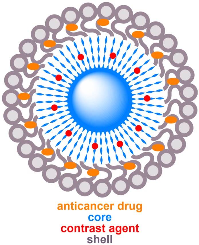
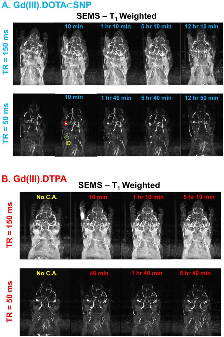
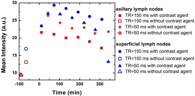
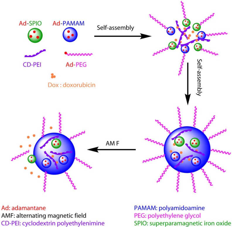
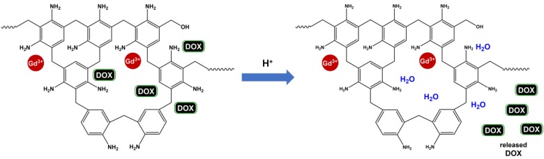
Cancer theranostics is one of the most important approaches for detecting and treating patients at an early stage. To develop such a technique, accurate detection, specific targeting, and controlled delivery are the key components. Various kinds of nanoparticles have been proposed and demonstrated as potential nanovehicles for cancer theranostics. Among them, polymer-like dendrimers and copolymer-based core-shell nanoparticles could potentially be the best possible choices. At present, magnetic resonance imaging (MRI) is widely used for clinical purposes and is generally considered the most convenient and noninvasive imaging modality. Superparamagnetic iron oxide (SPIO) and gadolinium (Gd)-based dendrimers are the major nanostructures that are currently being investigated as nanovehicles for cancer theranostics using MRI. These structures are capable of specific targeting of tumors as well as controlled drug or gene delivery to tumor sites using pH, temperature, or alternating magnetic field (AMF)-controlled mechanisms. Recently, Gd-based pseudo-porous polymer-dendrimer supramolecular nanoparticles have shown 4-fold higher T1 relaxivity along with highly efficient AMF-guided drug release properties. Core-shell copolymer-based nanovehicles are an equally attractive alternative for designing contrast agents and for delivering anti-cancer drugs. Various copolymer materials could be used as core and shell components to provide biostability, modifiable surface properties, and even adjustable imaging contrast enhancement. Recent advances and challenges in MRI cancer theranostics using dendrimer- and copolymer-based nanovehicles have been summarized in this review article, along with new unpublished research results from our laboratories.

Keywords: cancer theranostics; copolymer nanoparticle; dendrimer nanoparticle; magnetic resonance.


Conflict of interest statement

Competing Interests: The authors have declared that no competing interest exists.

Figures

Figure 1
Figure 1
Schematic diagram of dendrimer, illustrating core (red), branching points (green), surface terminal groups (blue), and position of encapsulated moiety (orange) that can be contrast agent or drug. The dotted spherical boundaries dictate the generation (G). G = 0 for the core, G = 1 for the next concentric shell, and so on. Nc represents the number of branches in the core, whereas Nb is the branch cell multiplicity. In this figure, Nc = 3 and Nb = 2, which gives the total number of surface terminal groups, Z = NcNbG = 48.
Figure 2
Figure 2
Schematic diagram of a typical core-shell structure formed by copolymers, illustrating core (blue), shell (grey), MRI contrast agent (red), and anticancer drug (orange).
Figure 3
Figure 3
Comparison of MR images of lymph nodes in mice. (A) MR images of lymph nodes after injection of Gd(III).DOTA⸦SNPs at different time points under time of retention (TR) 150 ms and 50 ms and time of echo (TE) 10 ms. Red, green, and yellow circles represent the brachial, axillary and superficial lymph nodes, respectively. (B) MR imaging of the lymph nodes after injection of Gd(III).DTPA at different time points with time of retention 150 ms and 50 ms, where (A) shows superior contrast at TR = 50 ms.
Figure 4
Figure 4
Plot of intensity vs. time after injection of SNP, which shows that the mean intensities of axillary and superficial nodes are highest 80 min after injecting Gd(III).DOTA⸦SNPs.
Figure 5
Figure 5
Schematic diagram of the theranostic application of a supramolecular nanoparticle assembly for induced hyperthermic treatment. The diagram shows that drugs (represented by small red balls) can be loaded during assembly of supramolecular nanoparticles, delivered to the tumor site, and released under AMF, which is controlled externally.
Figure 6
Figure 6
pH-controllable drug delivery and contrast-enhanced MR imaging based on core-shell smart polymer nanoparticles. At low pH, protonation of the amino groups on the shell leads to release of DOX molecules into the bulk solution. Upon release, the empty shell scaffolds take up water molecules, which interact with the inner-layer Gd, enhancing MRI contrast.
Figure 7
Figure 7
MR analysis of human cervical tumor-bearing mice using UCS-Gd-DOX. (A) 2D-slice images of tumor (black circles), where the color bar indicates the MR signal intensity. Only the image pixels with intensity values between ±2 standard deviations (STD) of the ROI signal were kept to avoid image artifacts. (B) SNR for each slice in (A) at time points: pre-scan (-206 min), 30 min, 82 min, 151 min, 203 min, 255 min, and 487 min. (C) Contrast-to-noise ratio (CNR) of tumor, liver, kidney, CuSO4, and noise area before and after injection. Here CNR % = (SNRpostinjection - SNRpreinjection) / SNRpreinjection ×100%. Error bars represent mean ± standard deviation (n = 3 mice). (D) T1-weighted MR images acquired by "spin-echo multiple slices" (SEMS) and then processed by "maximum intensity projection". Repetition time (TR) = 200 ms, echo time (TE) = 10 ms, field of view (FOV) = 64×32 mm2, matrix size = 256×128, slice thickness = 0.5 mm. The ROIs of tumor, liver, kidney, and CuSO4 reference are indicated by red, blue, green, and cyan shades, respectively.

References

    1. Janib SM, Moses AS, MacKay JA. Imaging and drug delivery using theranostic nanoparticles. Adv Drug Deliv Rev. 2010;62:1052–1063. - PMC - PubMed
    1. Sunderland CJ, Steiert M, Talmadge JE. et al. Targeted nanoparticles for detecting and treating cancer. Drug Dev Res. 2006;67:70–93.
    1. Kelkar SS, Reineke TM. Theranostics: Combining imaging and therapy. Bioconjug Chem. 2011;22:1879–1903. - PubMed
    1. Key J, Leary JF. Nanoparticles for multimodal in vivo imaging in nanomedicine. Int J Nanomedicine. 2014;9:711–726. - PMC - PubMed
    1. Huang G, Chen H, Dong Y. et al. Superparamagnetic iron oxide nanoparticles: Amplifying ros stress to improve anticancer drug efficacy. Theranostics. 2013;3:116–126. - PMC - PubMed

Publication types

MeSH terms