Review
. 2018 Dec 20:7:F1000 Faculty Rev-1960.
doi: 10.12688/f1000research.16451.1. eCollection 2018.

Oscillations and Spike Entrainment

Affiliations
Review

Oscillations and Spike Entrainment

Charles J Wilson et al. F1000Res. .

Abstract

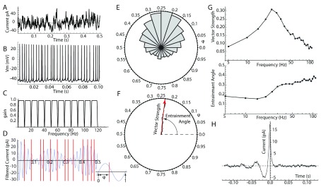
Oscillatory input to networks, as indicated by field potentials, must entrain neuronal firing to be a causal agent in brain activity. Even when the oscillatory input is prominent, entrainment of firing is not a foregone conclusion but depends on the intrinsic dynamics of the postsynaptic neurons, including cell type-specific resonances, and background firing rates. Within any local network of neurons, only a subset of neurons may have their firing entrained by an oscillating synaptic input, and oscillations of different frequency may engage separate subsets of neurons.

Keywords: LFP; oscillation; phase-locking.


Conflict of interest statement

No competing interests were disclosed.No competing interests were disclosed.No competing interests were disclosed.

Figures

Figure 1.
Figure 1.. Measuring entrainment.
( A) Broadband noise current stimulus applied to the cell via intracellular (perforated patch) recording. ( B) Membrane potential responses and spiking in response to the broadband stimulus. ( C) Series of bandpass filters used to separate frequency components of the current stimulus. ( D) One of the filtered input frequency components (25 Hz; blue waveform) and spike times (red lines). Inset: Measurement of spike phase (φ) from the filtered frequency component. ( E) Phase histogram for spikes measured on the 25 Hz frequency component. ( F) Normalized vector sum of all the spike phases from the histogram in E. Length of the result is the vector strength, and the angle of the resultant vector is the entrainment angle. ( G) Vector strength (upper) and entrainment angle (lower) for each of the frequency components obtained using the filter set shown in C. ( H) Spike-triggered average of the same data used in AG. Note oscillation at about 25 Hz, corresponding to the peak in the entrainment spectrum shown in G.
Figure 2.
Figure 2.. Measuring membrane resonance.
( A) Voltage clamp protocol for measuring resonance. The neuron’s voltage is clamped to a sine wave voltage chirp (middle) centered on a holding potential ( H). The frequency of the sine wave is increased linearly from 0 to 40 Hz over 40 seconds (bottom). The amplitude of the resulting sinusoidal clamp current (top) is inversely proportional to the membrane impedance. The minimum current amplitude around 11 s indicates the resonant frequency for the cell’s subthreshold impedance. ( B) Impedance, measured as the ratio of voltage command sine wave amplitude to clamp current, over the range of frequencies in the voltage command chirp. The impedance peaks at a resonant frequency near 11 Hz. ( C) The phase angle (angle between the voltage command and the current) at each frequency. At low frequencies, the voltage leads the current, whereas at high frequencies beyond the cell’s resonant frequency, the voltage lags behind the current. The frequency of zero phase difference occurs near but slightly below the frequency of peak resonance.
Figure 3.
Figure 3.. Entrainment modes.
Neuronal membrane potential is shown in blue, and stimulus current waveform is overlaid in red. ( A) Rate modulation occurs when the cell’s firing rate is faster than the stimulus frequency. The cell’s background firing rate is 15 spikes/s. The stimulus frequency is 1 Hz. ( B) One-to-one phase-lock occurs when the stimulus frequency is near the cell’s firing rate. Stimulus frequency is 15 Hz. ( C) Irregular firing occurs when the stimulus frequency and the cell firing rate are not commensurable.
Figure 4.
Figure 4.. Frequency-dependent spike entrainment in a non-resonant striatal spiny neuron.
Two different frequencies of sinusoidal current (26 and 45 Hz) were injected intracellularly while the cell was firing at one of two frequencies: 26 or 45 spikes/s. The firing times are compared with the membrane voltage waveform when the cell was hyperpolarized slightly to prevent firing and receiving the same two sinusoids or each of the sinusoidal currents was presented alone. ( A) Firing at 26 spikes/s while stimulated with both sine waves. ( B) The subthreshold membrane potential waveform for the same cell with the same stimulus but hyperpolarized slightly to prevent spikes. Note that the action potentials shown in A do not align on the voltage peak or any other consistent feature of the membrane potential waveform. ( C) The subthreshold membrane potential response to the 26 Hz sinusoidal current presented alone. Note that spikes generated in the presence of both frequencies are phase-locked to the 26 Hz sine wave component, regardless of the presence of the 45 Hz sine wave. ( D) The same neuron in the presence of the same stimulus shown in A but now firing at 45 spikes/s. ( E) Spike timing at 45 spikes/s compared with the subthreshold membrane potential waveform. Again, spikes are not generated at the times of peak depolarization by the two-frequency stimulus. ( F) Spike timing when both stimuli are present, compared with the subthreshold membrane potential waveform in the presence of the 45 Hz sine wave alone. Note that, in each case, the neuron is phase-locked to the frequency component close to its own firing rate and there is little influence from the other stimulus frequency.

References

    1. Adrian Ed, Matthews BHC: The Berger rhythm: potential changes from the occipital lobes in man. Brain. 1934;57(4):355–85. 10.1093/brain/57.4.355 - DOI - PubMed
    1. Buzsáki G: Rhythms of the Brain. Oxford University Press;2006. 10.1093/acprof:oso/9780195301069.001.0001 - DOI
    1. Fries P: Rhythms for Cognition: Communication through Coherence. Neuron. 2015;88(1):220–35. 10.1016/j.neuron.2015.09.034 - DOI - PMC - PubMed
    2. F1000 Recommendation

    1. Jefferys JG: Nonsynaptic modulation of neuronal activity in the brain: electric currents and extracellular ions. Physiol Rev. 1995;75(4):689–723. 10.1152/physrev.1995.75.4.689 - DOI - PubMed
    1. Buzsáki G, Anastassiou CA, Koch C: The origin of extracellular fields and currents--EEG, ECoG, LFP and spikes. Nat Rev Neurosci. 2012;13(6):407–20. 10.1038/nrn3241 - DOI - PMC - PubMed
    2. F1000 Recommendation

Publication types

MeSH terms

LinkOut - more resources