. 2018 Nov;2(10):314-323.
doi: 10.4049/immunohorizons.1800049.

The Neuropeptides of Ocular Immune Privilege, α-MSH and NPY, Suppress Phagosome Maturation in Macrophages

Affiliations

The Neuropeptides of Ocular Immune Privilege, α-MSH and NPY, Suppress Phagosome Maturation in Macrophages

Isaac J Benque et al. Immunohorizons. 2018 Nov.

Abstract

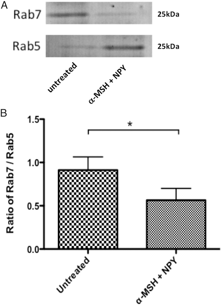
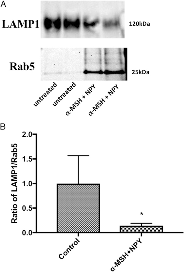
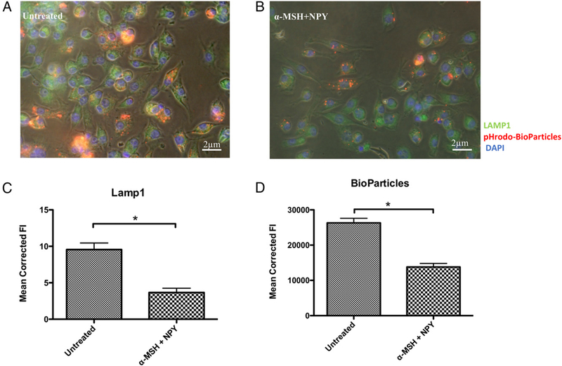
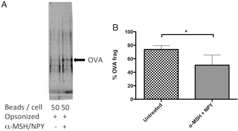
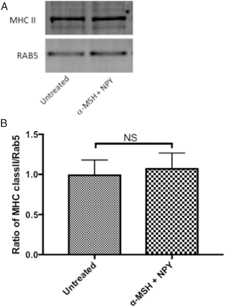
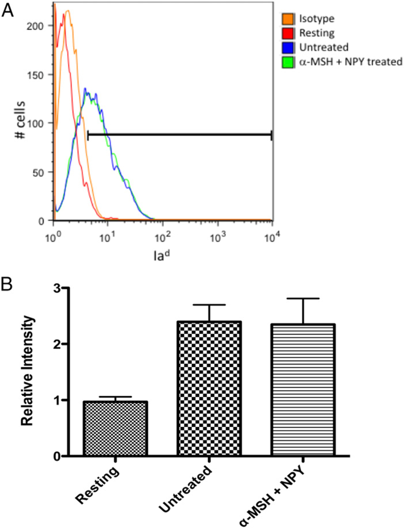
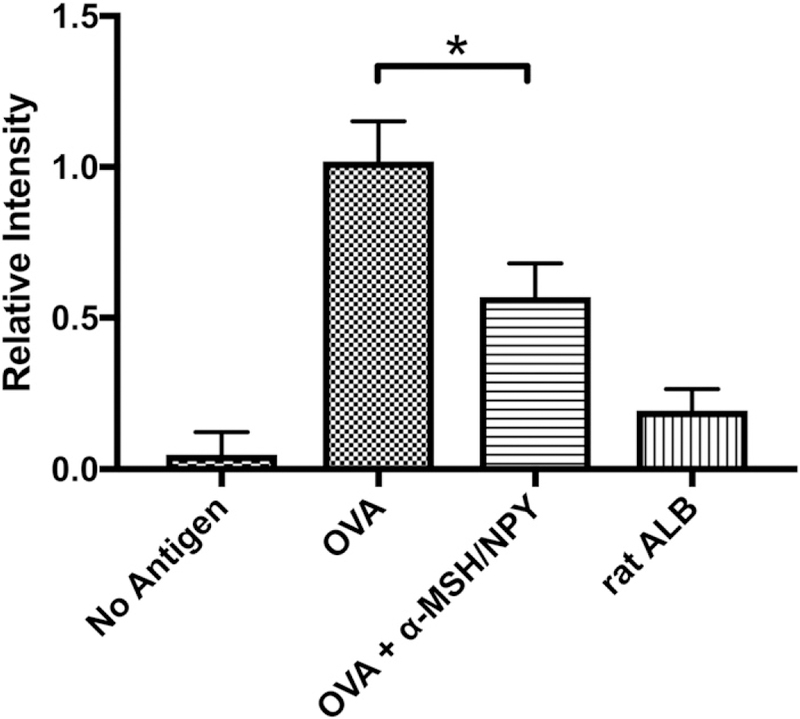
The ocular microenvironment has evolutionarily adapted several mechanisms of immunosuppression to minimize the induction of inflammation. Neuropeptides produced by the retinal pigment epithelial cells regulate macrophage activity. Two neuropeptides, α-melanocyte-stimulating hormone (α -MSH) and neuropeptide Y (NPY), are constitutively expressed by the retinal pigment epithelial cells. Together these two neuropeptides induce anti-inflammatory cytokine production in endotoxin-stimulated macrophages and suppress phagocytosis of unopsonized bioparticles. These neuropeptides do not suppress the phagocytosis of opsonized bioparticles; however, they do suppress phagolysosome activation or formation. In this report, we studied the possibility that α-MSH with NPY suppress phagosome maturation within macrophages using opsonized OVA-coated magnetic beads to isolate and analyze the phagosomes. The magnetic bead-containing intercellular vesicles were isolated and assayed for Rab5, Rab7, LAMP1, Iad, and OVA. The macrophages cotreated with α-MSH and NPY were suppressed in Rab7 recruitment to the phagosome with suppression in LAMP1 expression but not in Iad expression. The results demonstrated that the α-MSH/NPY cotreatment suppressed phagosome maturation. In addition, the a-MSH/NPY-cotreated macrophages were suppressed in their ability to Ag stimulate CD4+ T cell proliferation. These results imply a potential mechanism of ocular immune privilege to divert Ag processing to prevent autoreactive effector T cells from binding their target cognate Ag within the ocular microenvironment.


Conflict of interest statement

DISCLOSURES A.W.T. is a consultant for Palatin Technologies, Cranbury, NJ. The other authors have no financial conflicts of interest.

Figures

FIGURE 1.
FIGURE 1.. The effects of neuropeptide treatment on phagosome maturation.
The isolated magnetic bead–containing vesicles were immunoblotted for Rab5 and Rab7. The ratio of Rab7 to Rab5 was calculated from the intensity of the bands. Presented are (A) a representative immunoblot and (B) the mean ± SEM of the relative ratio of Rab7/Rab5 from three independent experiments. *There is a highly significant (p = 0.008) suppression in the expression of Rab7 to Rab5 in the macrophages cotreated with α-MSH and NPY. This demonstrates suppression in the maturation of the phagosomes.
FIGURE 2.
FIGURE 2.. The effects of neuropeptide treatment on phagolysosome formation.
The isolated magnetic bead–containing vesicles were immunoblotted for Rab5 and LAMP1. The ratio of LAMP1 to Rab5 was calculated from the intensity of the bands. Presented are (A) a representative immunoblot and (B) the mean 6 SEM of the relative ratio of LAMP1/Rab5 from four independent experiments. *There is a significant (p = 0.03) suppression in the expression of LAMP1 to Rab5 in the macrophages cotreated with α-MSH and NPY. This demonstrates suppression in the formation of phagolysosomes.
FIGURE 3.
FIGURE 3.. The effects of neuropeptide treatment on LAMP1 expression.
The macrophages were fed opsonized pHrodo–S. aureus bioparticles, fixed, and stained for LAMP1 and DAPI. Representative images of (A) untreated and (B) α-MSH/NPY–cotreated macrophages. (C and D) The bar graphs are the mean fluorescent intensities ± SEM of 15 independent experiments. This demonstrates a suppression in LAMP1 protein production in the treated macrophages. *There was a highly significant (p ± 0.001) suppression of LAMP1 expression (C) in the α-MSH/NPY–cotreated macrophages, with a corresponding suppression in pHrodo Red BioParticle intensity (D).
FIGURE 4.
FIGURE 4.. Effects of α -MSH/NPY cotreatment on OVA fragmentation within macrophages.
(A) The isolated magnetic bead–containing vesicles were immunoblotted for OVA. (B) The percentage of OVA fragmented was calculated by the ratio of the intensities of OVA fragments over the sum of the fragments plus whole OVA band intensities. Significant suppression of OVA frag-mentation was detected in macrophages cotreated with α-MSH and NPY. Presented are the mean ± SEM of the percent OVA fragments (% OVA frag) of three independent experiments, with *p ≤ 0.01.
FIGURE 5.
FIGURE 5.. The effects of neuropeptide treatment on MHC II within magnetic bead–containing vesicles.
The isolated magnetic bead–containing vesicles were immunoblotted for Rab5 and MHC II. The ratio of MHC II to Rab5 was calculated from the intensity of the bands. Presented are (A) representative immunoblot and (B) the mean ± SEM of the relative ratio of MHC II/Rab5 from three independent experiments. There was no significant suppression in the expression of MHC II to Rab5 in the macrophages cotreated with α-MSH and NPY. This demonstrates that MHC II is still associated with phagosomes.
FIGURE 6.
FIGURE 6.. Effects of neuropeptide treatment on Iad expression on macrophages.
The α-MSH/NPY–cotreated macrophages were fixed and stained for surface expression of Iad and analyzed by flow cytometry. (A) Repre-sentative histogram of three independent experiments with the gate set to exclude ˃.95% of isotype-stained cells. (B) The bar graph presents the relative intensity of Iad expression on untreated and α-MSH/NPY– cotreated macrophages given opsonized nonfluorescent bioparticles to phagocytize, and resting macrophages. No significant difference was seen between activated macrophages treated or untreated in Iad ex-pression. This demonstrates that MHC II expression is not altered in the treated macrophages.
FIGURE 7.
FIGURE 7.. Effects of neuropeptide treatment on APC stimulation of Ag-specific T cells.
The α-MSH/NPY–cotreated macrophages were fed opsonized OVA to process. OVA-specific T cells were added to the wells and assayed for proliferation. Presented are the relative levels of proliferation in comparison with the proliferation detected in the cultures of untreated OVA-presenting APC with OVA-specific T cells (OVA). Wells of no Ag contained only cultured APC with OVA-specific T cells. For Ag specificity, opsonized rat albumin was used instead of opsonized OVA (ratALB). The results presented are from nine independent cultures. This demonstrates that the treated APC are suppressed in their ability to stimulate Ag-specific T cell proliferation. The neuropeptide-treated APC were significantly suppressed (*p ≤ 0.001) in their ability to stimulate T cell proliferation.

References

    1. Taylor AW, and Ng TF. 2018. Negative regulators that mediate ocular immune privilege. J. Leukoc. Biol 103: 1179–1187. - PMC - PubMed
    1. Lau CH, and Taylor AW. 2009. The immune privileged retina mediates an alternative activation of J774A.1 cells. Ocul. Immunol. Inflamm 17: 380–389. - PMC - PubMed
    1. Kawanaka N, and Taylor AW. 2011. Localized retinal neuropeptide regulation of macrophage and microglial cell functionality. J. Neuro- immunol 232: 17–25. - PMC - PubMed
    1. Hsu SM, Mathew R, Taylor AW, and Stein-Streilein J. 2014. Ex- vivo tolerogenic F4/80+ antigen-presenting cells (APC) induce efferent CD8+ regulatory T cell-dependent suppression of experimental autoimmune uveitis. Clin. Exp. Immunol 176: 37–48. - PMC - PubMed
    1. Taylor AW, Dixit S, and Yu J. 2015. Retinal pigment epithelial cell line suppression of phagolysosome activation. Int. J. Ophthalmol. Eye Sci DOI: 10.19070/2332-290X-SI02001. - DOI - PMC - PubMed