. 2018 Dec 11:10.1111/nph.15630.
doi: 10.1111/nph.15630. Online ahead of print.

The Selaginella rhizophore has a unique transcriptional identity compared with root and shoot meristems

Affiliations

The Selaginella rhizophore has a unique transcriptional identity compared with root and shoot meristems

Alison Mello et al. New Phytol. .

Abstract

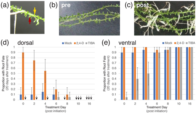
The genus Selaginella resides in an early branch of the land plant lineage that possesses a vasculature and roots. The majority of the Selaginella root system is shoot borne and emerges through a distinctive structure known as the rhizophore, the organ identity of which has been a long-debated question. The rhizophore of Selaginella moellendorffii - a model for the lycophytes - shows plasticity to develop into a root or shoot up until 8 d after angle meristem emergence, after which it is committed to root fate. We subsequently use morphology and plasticity to define the stage of rhizophore identity. Transcriptomic analysis of the rhizophore during its plastic stage reveals that, despite some resemblance to the root meristem, rhizophore gene expression patterns are largely distinct from both shoot and root meristems. Based on this transcriptomic analysis and on historical anatomical work, we conclude that the rhizophore is a distinct organ with unique features.

Keywords: Selaginella moellendorffii; early meristem; lycophyte evolution; lycophyte roots; rhizophore.


Figures

Fig. 1
Fig. 1
The shoot-borne rhizophores of Selaginella moellendorffii transition to roots. The majority of the S. moellendorffii root system is derived from outgrowths in the shoot (a). A ventral meristem becomes a rhizophore (arrow) that transitions to a root (b). This root system expands by successive, bifurcations at the root apex (c). Dorsal angle meristems typically give rise to shoots, which maintain an apical cell (arrow) at the surface of the meristem (d). Rhizophores transition to roots when the apical cell becomes internal giving rise to a nascent cap in early transition stages (e) that forms a multilayered cap at later stages (f). The root in (e) is in the process of transitioning from rhizophore and still lacked root hairs (not shown) while the root in (f) had root hairs and a well-defined internal apical cell (arrow). Scale bars are 25 µm.
Fig. 2
Fig. 2
Window of developmental plasticity in Selaginella moellendorffii exhibited through auxin treatment or perturbation. Developing dorsal or ventral angle meristem outgrowths at various developmental stages (0,2,4,6,8,10,16 days post initiation) were treated with mock solution (blue), 10µM 2,4-D (orange) or 100µM TIBA (gray) and organ identities (root or shoot) were assayed 20 days post treatment. The location of application of the lanolin paste is shown in (a). Red arrow indicates angle meristem location (shoot angle); yellow arrow, the site treatment was applied (on ventral surface). Comparison of overall morphology of pre-treatment (b) vs 2,4-D treated (c) samples shows that auxin treatment greatly enhances root density. Quantitative analysis of the results of either mock, 2,4-D, or TIBA treatment on the dorsal (c) and ventral meristems (d). For each developmental stage, 20 angle meristems outgrowths not yet showing shoot or root features were treated. All outgrowths that did not show root fate by day 20 adopted shoot fates. Error bars show standard error of the proportion at 95% confidence limits. Black arrows indicate a frequency of zero.
Fig. 3
Fig. 3
The rhizophore shows a distinct transcriptional signature with some affinity to root. (a) Row normalized mean expression values of replicate samples showing significantly differentially expressed genes across all sampled tissues. (b) Multi-dimensional scaling of transcriptional profile of root, rhizophore, shoot meristem and leaves (performed in R using the cmdscale function). Note that leaf and shoot are merged into one point because they could not be distinguished at the scale of the figure. (c) Venn diagram of the single- or paired- meristem upregulated transcripts in the root, shoot, and shoot meristematic zones. Only genes with mean RPKM values >0.1 were counted. Color key represents row normalized Z-scores.
Fig. 4
Fig. 4
Heatmaps of meristem-specific and meristem-shared upregulated gene sets show distinct transcriptional programs of shoot and rhizophore. Shoot (a), rhizophore (b), and root (c) specific genes were identified as those showing significant upregulation in one meristem compared to both other meristems. Root-Rhizophore (d), Root-Shoot (e), and Rhizophore-Shoot (f) genes were identified by showing significant upregulation in two meristems compared to the third (see the Materials and Methods section). Note that leaf samples are shown but were left out of the comparisons because analysis shows they are highly similar to the shoot meristem. Color key represents row normalized Z-scores.
Fig. 5
Fig. 5
Gene ontology (GO)-term enrichment analysis of meristem-specific and meristem-shared genes. Color coding represents P-value of the term’s enrichment in a meristem-specific or meristem-shared transcript list, where red<0.001; orange<0.01; blue<0.05. Note that terms scoring pval>0.03 are not listed for space considerations. See Supporting Information Table S6 for complete list and other information.
Fig. 6
Fig. 6
DR5 Reporter is highly induced in Selaginella moellendorffii root cells. Protoplasts derived from whole S. moellendorffii root tissue transiently transfected with the DR5::GFP were incubated for 18 hours after transfection. The same batch of transfected protoplasts were split and then subjected 10µM 2,4-D or a mock treatment. The two treatments, mock (left) or 2,4 D (right), were visualized under the same excitation and exposure settings six hours after the treatments.

References

    1. Achard P, Cheng H, De Grauwe L, Decat J, Schoutteten H, Moritz T, Van Der Straeten D, Peng J, Harberd NP. 2006. Integration of plant responses to environmentally activated phytohormonal signals. Science 311(5757): 91–94. - PubMed
    1. Aida M, Beis D, Heidstra R, Willemsen V, Blilou I, Galinha C, Nussaume L, Noh Y-S, Amasino R, Scheres B. 2004. The PLETHORA genes mediate patterning of the Arabidopsis root stem cell niche. Cell 119(1): 109–120. - PubMed
    1. Banks JA. 2009. Selaginella and 400 million years of separation. Annu Rev Plant Biol 60: 223–238. - PubMed
    1. Banks JA. 2015. The evolution of the shoot apical meristem from a gene expression perspective. New Phytol 207(3): 486–487. - PubMed
    1. Banks JA, Nishiyama T, Hasebe M, Bowman JL, Gribskov M, dePamphilis C, Albert VA, Aono N, Aoyama T, Ambrose BA, et al. 2011. The Selaginella genome identifies genetic changes associated with the evolution of vascular plants. Science 332(6032): 960–963. - PMC - PubMed

Associated data