. 2020 Jan;51(1):34-46.
doi: 10.1111/ejn.14334. Epub 2019 Jan 23.

Circadian rhythm of redox state regulates membrane excitability in hippocampal CA1 neurons

Affiliations

Circadian rhythm of redox state regulates membrane excitability in hippocampal CA1 neurons

Ghazal Naseri Kouzehgarani et al. Eur J Neurosci. 2020 Jan.

Abstract

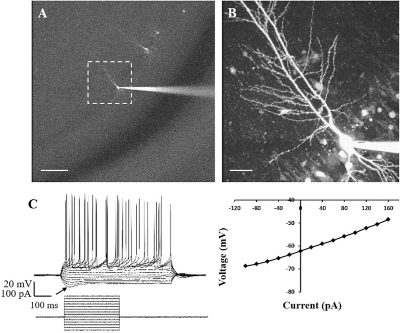
Behaviors, such as sleeping, foraging, and learning, are controlled by different regions of the rat brain, yet they occur rhythmically over the course of day and night. They are aligned adaptively with the day-night cycle by an endogenous circadian clock in the suprachiasmatic nucleus (SCN), but local mechanisms of rhythmic control are not established. The SCN expresses a ~24-hr oscillation in reduction-oxidation that modulates its own neuronal excitability. Could circadian redox oscillations control neuronal excitability elsewhere in the brain? We focused on the CA1 region of the rat hippocampus, which is known for integrating information as memories and where clock gene expression undergoes a circadian oscillation that is in anti-phase to the SCN. Evaluating long-term imaging of endogenous redox couples and biochemical determination of glutathiolation levels, we observed oscillations with a ~24 hr period that is 180° out-of-phase to the SCN. Excitability of CA1 pyramidal neurons, primary hippocampal projection neurons, also exhibits a rhythm in resting membrane potential that is circadian time-dependent and opposite from that of the SCN. The reducing reagent glutathione rapidly and reversibly depolarized the resting membrane potential of CA1 neurons; the magnitude is time-of-day-dependent and, again, opposite from the SCN. These findings extend circadian redox regulation of neuronal excitability from the SCN to the hippocampus. Insights into this system contribute to understanding hippocampal circadian processes, such as learning and memory, seizure susceptibility, and memory loss with aging.

Keywords: CA1 pyramidal neurons; circadian clock; rat hippocampus; reduction-oxidation; suprachiasmatic nucleus.


Conflict of interest statement

COMPETING INTERESTS

The authors have no conflict of interest to declare.

Figures

Figure 1.
Figure 1.. The hippocampal CA1 region and the SCN of the same animal exhibit opposite redox states in day vs. night.
During the subjective day (CT 6), the SCN is more reduced and the CA1 is more oxidized, while during the subjective night (CT 14) the SCN is more oxidized and the CA1 is more reduced. (A) The SCN shows low BioGEE incorporation at CT 6 compared with high BioGEE incorporation at CT 14, reflecting a reduced state at CT 6 and oxidized state at CT 14. Scale bar = 500 μm. (B) Conversely, the hippocampal CA1 area shows high BioGEE incorporation at CT 6 and low BioGEE incorporation at CT 14, reflecting an oxidized state at CT 6 and reduced state at CT 14. Scale bar = 500 μm. (C) The intensity of DAB staining of BioGEE was measured using ImageJ. Representative images from CT 6 are shown with regions of interest (ROIs) around the SCN and the hippocampal CA1 layer (solid line) and control regions (dashed line). DAB staining intensity in the SCN is significantly higher at CT 14 vs. CT 6 (P = 0.01), but in the CAI layer of hippocampus, staining is higher at CT 6 vs. CT 14 (P = 0.03). DAB intensity levels were significantly lower in the hippocampal CA1 region at CT 6 and CT 14 vs. the SCN at CT 14 (P = 0.01 and 0.004, respectively). Two-way mixed-model ANOVA; Tukey’s post-hoc test. *P < 0.05, **P < 0.01, n = 3. 3V = 3rd Ventricle, OC = Optic Chiasm. Scale bar = 500 μm. (D) Quantification of western blots for comparison of endogenous biotin in rat SCN and hippocampal slices incubated with EBSS at CT 6 and CT 14 showed no statistically significant differences between the two time points (SCN: CT 6 (87.64 ± 4.18 %) vs. CT 14 (84.32 ± 5.78 %), P = 0.65; hippocampus: CT 6 (79.50 ± 4.19 %) vs. CT 14 (66.63 ± 3.72 %), P = 0.08). Student’s unpaired t-test, n = 3–5. ns = non-significant. Error bars represent SEM.
Figure 2.
Figure 2.. The hippocampal CA1 layer undergoes an endogenous oscillation in redox state with a ~24-h period.
(A) Excised hippocampal tissue stained with DAPI to label cell nuclei displays the tri-synaptic loop of information processing: information enters through the dentate gyrus (DG), passes through the CA3 layer, and leaves via the CA1 region. Scale bar = 500 μm. (B) Ratiometric imaging of the endogenous fluorescence of FAD/NAD(P)H was performed on the CA1 region of the live hippocampal slice. Excitation wavelength was set to 730 nm and two windows of emission at 430–500 nm and 500–550 nm were recorded simultaneously. Scale bar = 100 μm. (C) Real-time imaging of the relative redox state in the CA1 region of hippocampal brain slices revealed a near-24-h oscillation over 72 h. Measurement was taken from the box in (B) over this period. Ratio of FAD (500+ nm)/NAD(P)H (400+ nm) plotted against time (h) shows an oscillation. Period (τ) = 22.75, determined by χ2 periodogram analysis. Long-term imaging of a second hippocampal slice over 72 hours recorded an oscillation over 36 hours that resembled the first, although the duration did not permit periodogram analysis.
Figure 3.
Figure 3.. Hippocampal CA1 pyramidal neurons exhibit distinctive morphological and electrophysiological properties using dye-filling and whole-cell patch clamp recording.
(A) Multiple pyramidal neurons along the CA1 layer were filled with Alexa Fluor 488 dye. Scale bar = 100 μm. (B) Image of a single CA1 pyramidal neuron shown in the dashed box in (A) was taken on a two-photon laser-scanning microscope. Scale bar = 20 μm. (C) Voltage response of a hippocampal CA1 pyramidal neuron was measured in response to hyperpolarizing and depolarizing current pulses ranging from −100 to +160 pA with 20 pA increments. The arrow points to the depolarizing sag in response to hyperpolarizing currents, indicating the hyperpolarization-activated cation current (Ih). n = 112, 112 brain slices, 30 animals.
Figure 4.
Figure 4.. Membrane excitability of hippocampal CA1 pyramidal neurons oscillates in vitro with neurons more depolarized in subjective late night/early subjective day and significantly hyperpolarized in subjective mid-day.
(A) Individual CA1 neurons (grey) and 2h running averages (black) exhibit oscillation in Vm assessed over 21 h in vitro (n = 112 cells in 112 brain slices from 30 animals). (B) Mean Vm of CA1 neurons over 6 CT intervals is most hyperpolarized at CT 7 (−72.73 ± 0.56 mV) and most depolarized at CT 1 (−68.29 ± 0.79 mV, P = 0.0002), CT 14 (−69.46 ± 0.79 mV, P = 0.01), and CT 21 (−66.06 ± 1.78 mV, P = 0.007). One-way ANOVA; Tukey’s post-hoc test. *P < 0.05, **P < 0.01, ***P < 0.001, n = 4–40/CT. Error bars represent SEM.
Figure 5.
Figure 5.. Membrane input resistance of hippocampal CA1 pyramidal neurons undergoes a time-of-day change in vitro.
(A) Individual CA1 neurons (grey) and 2-h running averages (black) exhibit a difference in Rin measured from I-V curves constructed by current steps from −100 to +200 pA with 20 pA increments and a 600 ms duration (n = 112). This is a measure of the initial resistance of the cell at the beginning of the recording. (B) Individual CA1 neurons (grey) and 2-h running averages (black) exhibit a change in Rin measured from hyperpolarizing current steps which assesses the health of the neuron and the stability of the patch throughout the recording (n = 112). (C) Average Rin measured from the slope of I-V curves at six CT intervals has significantly lower input resistances at CT 11 (90.04 ± 9.51 MΩ) vs. CT 1 (115.27 ± 7.96 MΩ, P = 0.04) and CT 21 (119.97 ± 13.45 MΩ, P = 0.04). (D) Average Rin from hyperpolarizing current steps at six CT intervals show significantly lower input resistances at CT 11 (115.22 ± 18.35 MΩ) vs. CT 14 (167.01 ± 17.17 MΩ, P = 0.04) and CT 21 (172.16 ± 25.96 MΩ, P = 0.04). One-way ANOVA; Tukey’s post-hoc test. *P < 0.05, n = 7–47/CT. Error bars represent SEM.
Figure 6.
Figure 6.. Redox-induced changes in membrane excitability of hippocampal CA1 pyramidal neurons align with the endogenous redox oscillation of the hippocampus.
(A) Glutathione (GSH, 1mM, 5 min) produced a rapid, reversible membrane depolarization from the baseline with magnitudes significantly larger during mid-day (CT 7) compared to the early night (CT 14). (B) GSH-induced depolarization in 63 individual CA1 pyramidal neurons showed oscillation over 21 h throughout the circadian cycle (n = 1–6/CT, total of 63 brain slices, 20 animals). (C) GSH-induced depolarization of Vm in CA1 neurons at five CT groups showed the smallest ΔVm at CT 14 (5.51 ± 0.78 mV) and the largest ΔVm at CT 1 (12.49 ± 1.69 mV, P = 0.003), CT 7 (10.66 ± 0.91 mV, P = 0.0006), and CT 20 (10.89 ± 1.02 mV, P = 0.0009). One-way ANOVA; Tukey’s post-hoc test. **P < 0.01, ***P < 0.001, n = 4–19/CT. (D) To assess the hippocampal intrinsic redox oscillation, quantification of all western blots assayed for BioGEE incorporation was performed. Tissue-level glutathiolation was lowest at CT 14 (28.06 ± 3.6 %) and highest at CT 2 (68.12 ± 11.61 %, P = 0.02), CT 6 (84.74 ± 8.9 %, P = 0.0009), CT 10 (90.15 ± 4.57 %, P = 0.0003), and CT 18 (75.33 ± 9.43 %, P = 0.005). This indicates that the hippocampus is most reduced at CT 14, which aligns with the smallest GSH-induced Vm change at this time point. One-way ANOVA; Tukey’s post-hoc test. *P < 0.05, **P < 0.01, ***P < 0.001, n = 4. Error bars represent SEM.

References

    1. Abe M, Herzog ED, Yamazaki S, Straume M, Tei H, Sakaki Y, Menaker M, Block GD. (2002) Circadian rhythms in isolated brain regions. J. Neurosci, 22: 350–356. - PMC - PubMed
    1. Andersen P, Bliss TV, Lomo T, Olsen LI, Skrede KK (1969) Lamellar organization of hippocampal excitatory pathways. Acta. Physiol. Scand, 76: 4A–5A. - PubMed
    1. Arszovski A, Borhegyi Z, Klausberger T (2014) Three axonal projection routes of individual pyramidal cells in the ventral CA1 hippocampus. Front. Neuroanat, 8: 53. - PMC - PubMed
    1. Bass J & Takahashi JS (2010) Circadian integration of metabolism and energetics. Science, 330: 1349–1354. - PMC - PubMed
    1. Besing RC, Rogers CO, Paul JR, Hablitz LM, Johnson RL, McMahon LL, Gamble KL (2017) GSK3 activity regulates rhythms in hippocampal clock gene expression and synaptic plasticity. Hippocampus, 27: 890–898. - PMC - PubMed

Publication types