. 2020 Jun;271(6):1174-1185.
doi: 10.1097/SLA.0000000000003053.

Staphylococcus aureus Biofilm Infection Compromises Wound Healing by Causing Deficiencies in Granulation Tissue Collagen

Affiliations

Staphylococcus aureus Biofilm Infection Compromises Wound Healing by Causing Deficiencies in Granulation Tissue Collagen

Sashwati Roy et al. Ann Surg. 2020 Jun.

Abstract

Objective: The objective of this work was to causatively link biofilm properties of bacterial infection to specific pathogenic mechanisms in wound healing.

Background: Staphylococcus aureus is one of the four most prevalent bacterial species identified in chronic wounds. Causatively linking wound pathology to biofilm properties of bacterial infection is challenging. Thus, isogenic mutant stains of S. aureus with varying degree of biofilm formation ability was studied in an established preclinical porcine model of wound biofilm infection.

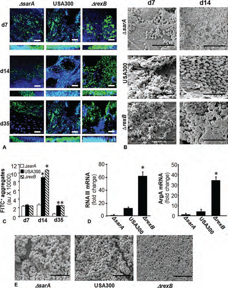
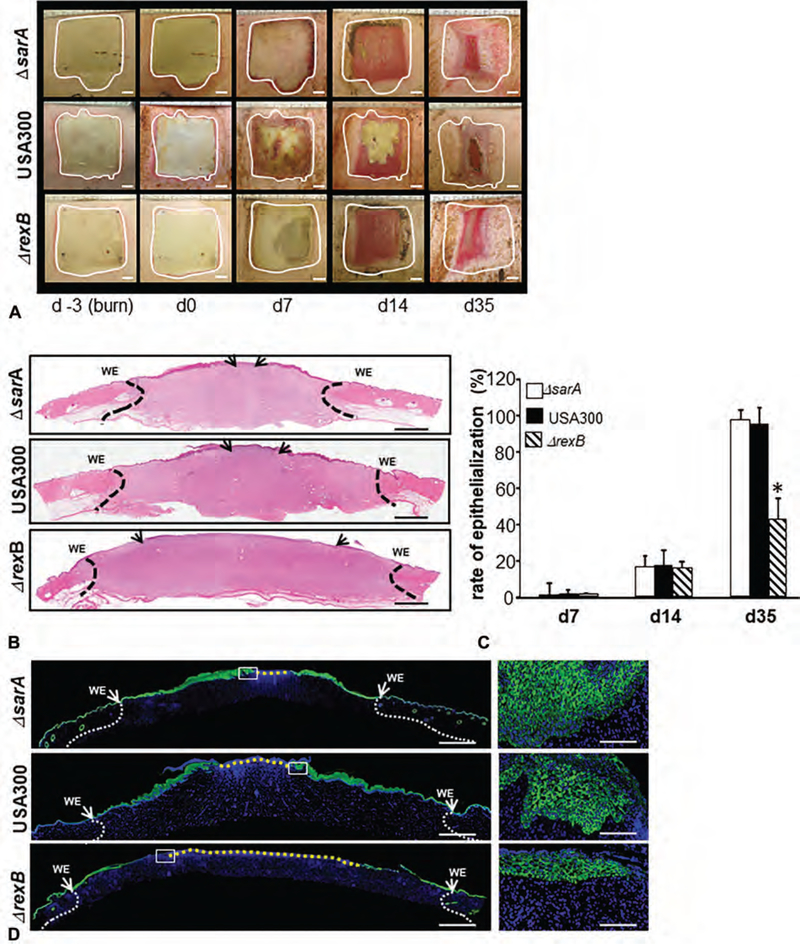
Methods: Isogenic mutant strains of S. aureus with varying degree (ΔrexB > USA300 > ΔsarA) of biofilm-forming ability were used to infect full-thickness porcine cutaneous wounds.

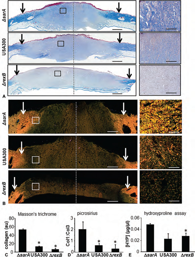
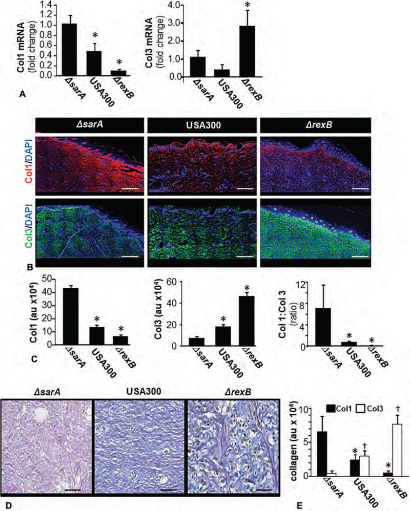
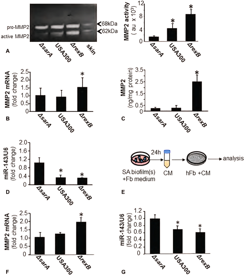
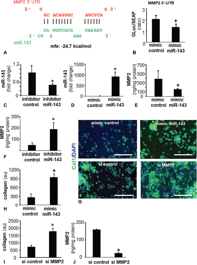
Results: Compared with that of ΔsarA infection, wound biofilm burden was significantly higher in response to ΔrexB or USA300 infection. Biofilm infection caused degradation of cutaneous collagen, specifically collagen 1 (Col1), with ΔrexB being most pathogenic in that regard. Biofilm infection of the wound repressed wound-edge miR-143 causing upregulation of its downstream target gene matrix metalloproteinase-2. Pathogenic rise of collagenolytic matrix metalloproteinase-2 in biofilm-infected wound-edge tissue sharply decreased collagen 1/collagen 3 ratio compromising the biomechanical properties of the repaired skin. Tensile strength of the biofilm infected skin was compromised supporting the notion that healed wounds with a history of biofilm infection are likely to recur.

Conclusion: This study provides maiden evidence that chronic S. aureus biofilm infection in wounds results in impaired granulation tissue collagen leading to compromised wound tissue biomechanics. Clinically, such compromise in tissue repair is likely to increase wound recidivism.


Conflict of interest statement

The authors report no conflicts of interest.

Figures

FIGURE 1.
FIGURE 1.
Evidence of hyperbiofilm formation by S. aureus, transposon mutant USA300::rexB while USA300::sarA mutant is a hypobiofilm-forming strain in porcine burn wound infections. Six 2 × 2 sq inch size burn wounds were created on back of pigs. On day 3 post-burn, the wounds were infected by isogenic strains of S. aureus USA300: USA300, USA300::rexB (ΔrexB) or USA300::sarA (ΔsarA). A, Representative confocal laser scanning microscopy images from burn wounds on days 7, 14, and 35 postinfection showing S. aureus (anti-SA, green) aggregates. Counterstaining was performed using DAPI (blue, nucleus). Z-stack images of tissues sections (20 μm) from burn wound are shown. Scale bar = 50 μm. B, Representative scanning electron microscopy (SEM) images from SA infected burn wounds on days 7 and 14 postinfection. Scale bar = 5 μm. C, Quantifications of S. aureus aggregates for images shown in (A). Data are mean ± SEM (n = 3), *P < 0.05 compared with ΔsarA. D, Quantification of mRNA levels of biofilm/virulence specific genes in biofilm infected burn wound tissue was determined using real-time PCR. The data was normalized against 16 s rRNA. Data are mean ± SEM (n = 6), *P < 0.05 compared with ΔsarA. E, SEM images of in vitro SA biofilms developed on polycarbonate filters. Scale bar = 5 μm.
FIGURE 2.
FIGURE 2.
Effects of S. aureus (SA) biofilm infection on burn wound healing evaluated using 3 strains of USA300 with varying degrees of biofilm-forming capability. On day 3 postburn, the wounds were infected by isogenic strains of S. aureus USA300, USA300::rexB (ΔrexB) or USA300::sarA (ΔsarA). A, Representative digital images of biofilm infected burn wounds on days 0, 7, 14, and 35 postinfection. The wound area on the day of burn has been demarcated with white dashed line. B, hematoxylin and eosin stained whole mount cross sections of infected burn wounds show re-epithelialization. The wound edges have been shown with dash black lines while the tip of migrated epithelium is shown with black arrows. WE indicates wound edge. Scale bar = 200 μm. C, rate of re-epithelialization (%) quantified from H&E stained images shown in (B). D, Immunofluorescence images of anti-K14 (green) stained biofilm infected burn wound tissue sections. Counter staining was performed with DAPI (blue, nucleus). Scale bar = 200 μm. Insets, zoom of the boxed area. Scale bar = 50 μm.
FIGURE 3.
FIGURE 3.
Hyperbiofilm infection by S. aureus USA::ΔrexB in burn wounds severely compromises granulation tissue collagen content. A–C, On day 3 postburn, the porcine wounds were infected by isogenic strains of S. aureus USA300, USA300::rexB (ΔrexB) or USA300::sarA (ΔsarA). A, Representative images of formalin-fixed paraffin-embedded (FFPE) biofilm infected day 35 burn wound biopsy sections (5 μm) were stained using Masson Trichrome (MT). MT staining results in blue-black nuclei, blue collagen, and light red or pink cytoplasm. Epidermal cells appear reddish. Scale bar = 200 μm. The edges of the wound have been shown with black/white arrows. Right panels are the zoom in images of the boxed areas within the images in the left panels. Scale bar, 50 μm. B, Representative images of formalin-fixed paraffin- embedded (FFPE) biofilm infected day 35 burn wound biopsy sections (5 μm) were stained using Picrosirius red (PS) staining. Scale bar = 200 μm. Insets zoom of the box area. Scale bar = 50 μm. C, Bar graph shows quantitation of collagen abundance using MT stains. Data are mean ± SEM (n = 6), *P < 0.05 compared with ΔsarA. D, Bar graph shows quantitation of collagen abundance using PS stains. Data are mean ± SEM (n = 3), *P < 0.05 compared with ΔsarA. E, Granulation (d35 postinfection) tissue collagen content was determined using hydroxyproline assay. Data are mean ± SEM (n = 6), *P < 0.05 compared with ΔsarA.
FIGURE 4.
FIGURE 4.
Dysregulation of Col1 and Col3 at wound-site granulation tissue in S. aureus biofilm-infected burn wounds. On day 3 post-burn, the wounds were infected by isogenic strains of S. aureus USA300, USA300::rexB (ΔrexB) or USA300::sarA (ΔsarA). Wound biopsies were collected at specified time-points after inoculation with S. aureus USA300 isogenic strains: USA300, USA300::rexB (ΔrexB), or USA300::sarA (ΔsarA). A, Expressions of Col1 and Col3 mRNA in wound biopsies collected on day 35 post-inoculation. Data presented as mean ± SEM (n = 4), * P < 0.05 compared with ΔsarA. B, Representative images of Col1 (red, anti-col1) and Col3 (green, anti-Col3) stained sections on days 35 post-inoculation. The sections were counterstained using DAPI (nuclear, blue). Scale bar = 100 μm. C, Bar graphs present quantitation of Col1 and Col3 signal intensity and their ratio in (B). Data are presented as mean ± SEM (n = 4), *P < 0.05 compared with ΔsarA. D, Herovici stained images of biofilm infected burn wound tissue sections from d35 post-infection burn wounds. Herovici stains young collagen reticulum blue (Col3) and mature collagen red (Col1) while providing a yellow cytoplasm counterstain. Nuclei are stained to black with Weigert Hematoxylin. E, Bar graphs present quantitation of Col1 and Col3 signal intensity and their ratio in (D). scale bar = 50 μm. Data are presented as mean ± SEM (n = 4), *P < 0.05 compared with Col1 of ΔsarA +P < 0.05 compared with Col3 of ΔsarA.
FIGURE 5.
FIGURE 5.
miR-143 and MMP-2 expression/activity in wounds fibroblasts is regulated by SA biofilm infection. Wound biopsies were collected at specified time points post-inoculation with S. aureus USA300 isogenic strains: USA300, USA300::rexB (ΔrexB) or USA300::sarA (ΔsarA). A, Representative, gelatin zymogram of d35 porcine infected burn wounds tissue. Based on molecular weight, pro- (70 kDa) and active MMP-2 (62 kDa) bands have been indicated. Quantification of active MMP-2 (62 kDa) band using densitometry. Data are mean ± SEM (n = 4) *P < 0.05 compared with ΔsarA. B, MMP-2 mRNA in d35 post-inoculation porcine infected burn wounds tissues quantified using real-time PCR. Data was normalized against 18S RNA as housekeeping gene. Data are mean ± SEM (n = 6) *P < 0.05 compared with ΔsarA. C, MMP-2 protein in d35 post-inoculation porcine infected burn wounds tissues quantified using ELISA. Data are mean ± SEM (n = 6). *P < 0.05 compared with ΔsarA. D, miR-143 expression in d35 post-inoculation porcine infected burn wounds tissues quantified using real-time PCR. Data was normalized against U6 snRNA as housekeeping genes. Data are mean ± SEM (n = 6) *P < 0.05 compared with ΔsarA. E-G, Human fibroblasts (hFb) were cultured with overnight conditioned media from SA biofilms. Mature biofilms developed on polycarbonate membrane in vitro were placed in cultured dish and incubated with fibroblast (Fb) culture media overnight. The conditioned media was harvested, centrifuged, filtered and then added to hFb. E, Schematic presentation of the experimental model. F, MMP-2 mRNA expression in hTERT immortalized cultured human fibroblasts (hFb) with conditioned media from biofilms cultured in vitro. Data are mean ± SEM (n = 6) *P < 0.05 compared with ΔsarA. G, miR-143 expression in hTERT immortalized cultured human fibroblasts (hFb) with conditioned media from biofilms cultured in vitro. Data are mean ± SEM (n = 3) *P < 0.05 compared with ΔsarA.
FIGURE 6.
FIGURE 6.
MMP-2 is a direct target of miR-143 and regulates fibroblast collagen levels. A, miR-143 predicted to target MMP-2 3’UTR based on RNA Hybrid algorithm. MMP-2 transcript is NM_004530. Binding position of miR-143 (green) corresponds to position 604–602 of 3′-UTR of MMP-2 (red). B, Luciferase activity in human fibroblasts after transfection with mimic miR-143 and mimic control. Data are mean ± SEM (n = 6) *P < 0.05 compared with mimic control. C and D, Expression of miR-143 in human fibroblasts transfected with (C) miR-143 inhibitor (D) miR-143 mimic. E and F, Expression of MMP-2 in human fibroblasts transfected with (E) miR-143 mimic (F) miR-143 inhibitor. G, Fluorescence microscopy images of collagen 1 (green) Col1 expression in hFb following bolstering of miR143 with mimic miR-143 (upper panel) and knockdown of MMP-2 with siMMP2. Counterstaining was performed using DAPI (blue, nuclear). H, Bar graph presents quantitation of Col1 from (G, upper panel). Data are mean ± SEM (n = 3). *P < 0.05 compared with mimic control. I–J, Knockdown of MMP-2 with siMMP2. Data are mean ± SEM (n = 4). *P < 0.05 compared with si control.
FIGURE 7.
FIGURE 7.
S. aureus biofilm results in loss of Col1 and reduces tensile strength through a miR-143-MMP-2-dependent pathway. Solid lines indicate pathways based on data from this work. Broken lines are based on literature (Eleswarapu SV et al, Tensile properties, collagen content, and crosslinks in connective tissues of the immature knee joint. PLoS One 2011; 6(10):e26178. Lodish H et al., Collagen: The Fibrous Proteins of the Matrix. In: Lodish H, Berk A, Zipursky Sea, eds. Molecular Cell Biology. New York: W. H. Freeman;2000. Roeder BA et al., Tensile mechanical properties of three-dimensional type I collagen extracellular matrices with varied microstructure. J Biomech Eng - Transactions of the Asme 2002; 124(2):214–222.

References

    1. Malone M, Bjarnsholt T, McBain AJ, et al. The prevalence of biofilms in chronic wounds: a systematic review and meta-analysis of published data. J Wound Care. 2017;26:20–25. - PubMed
    1. Wolcott R Disrupting the biofilm matrix improves wound healing outcomes. J Wound Care. 2015;24:366–371. - PubMed
    1. Percival SL, McCarty SM, Lipsky B. Biofilms and wounds: an overview of the evidence. Adv Wound Care (New Rochelle). 2015;4:373–381. - PMC - PubMed
    1. Barki KG, Das A, Dixith S, et al. Electric field based dressing disrupts mixed-species bacterial biofilm infection and restores functional wound healing. Ann Surg. 2017. Epub ahead of print. - PMC - PubMed
    1. Roy S, Elgharably H, Sinha M, et al. Mixed-species biofilm compromises wound healing by disrupting epidermal barrier function. J Pathol. 2014;233:331–343. - PMC - PubMed

Publication types