. 2019 Jun 1;28(11):1755-1767.
doi: 10.1093/hmg/ddz003.

Homozygous hydroxymethylbilane synthase knock-in mice provide pathogenic insights into the severe neurological impairments present in human homozygous dominant acute intermittent porphyria

Affiliations

Homozygous hydroxymethylbilane synthase knock-in mice provide pathogenic insights into the severe neurological impairments present in human homozygous dominant acute intermittent porphyria

Makiko Yasuda et al. Hum Mol Genet. .

Abstract

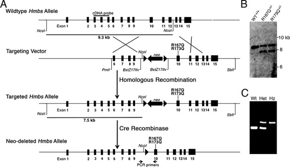
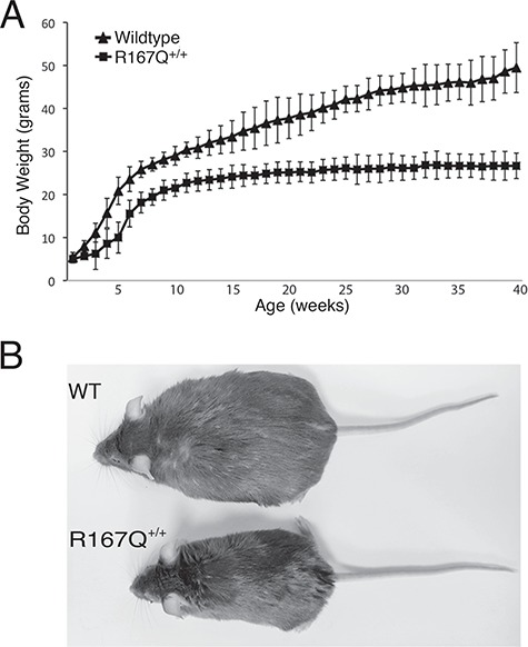
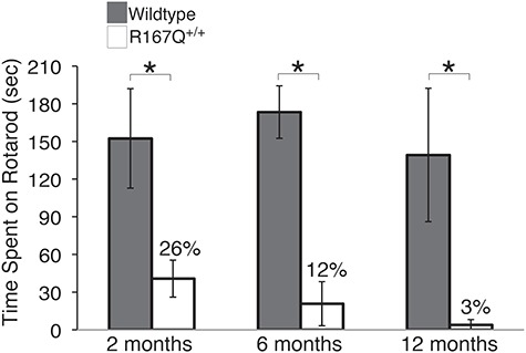
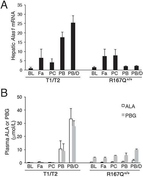
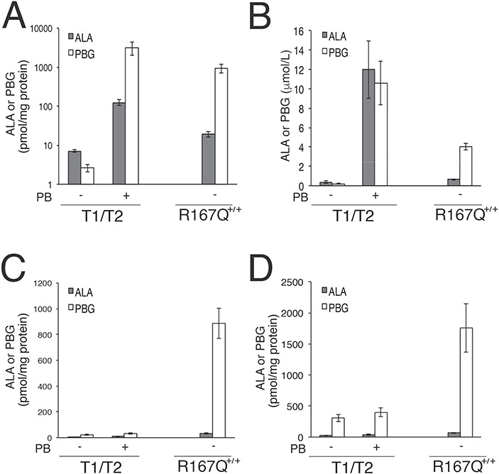
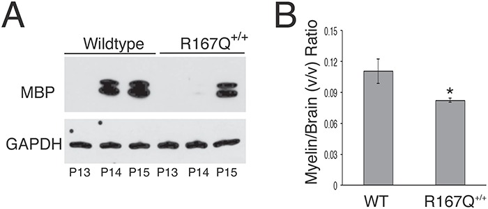
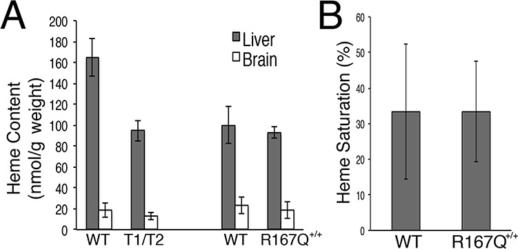
Acute intermittent porphyria (AIP) is an inborn error of heme biosynthesis due to the deficiency of hydroxymethylbilane synthase (HMBS) activity. Human AIP heterozygotes have episodic acute neurovisceral attacks that typically start after puberty, whereas patients with homozygous dominant AIP (HD-AIP) have early-onset chronic neurological impairment, including ataxia and psychomotor retardation. To investigate the dramatically different manifestations, knock-in mice with human HD-AIP missense mutations c.500G>A (p.Arg167Glu) or c.518_519GC>AG (p.Arg173Glu), designated R167Q or R173Q mice, respectively, were generated and compared with the previously established T1/T2 mice with ~30% residual HMBS activity and the heterozygous AIP phenotype. Homozygous R173Q mice were embryonic lethal, while R167Q homozygous mice (R167Q+/+) had ~5% of normal HMBS activity, constitutively elevated plasma and urinary 5-aminolevulinic acid (ALA) and porphobilinogen (PBG), profound early-onset ataxia, delayed motor development and markedly impaired rotarod performance. Central nervous system (CNS) histology was grossly intact, but CNS myelination was delayed and overall myelin volume was decreased. Heme concentrations in liver and brain were similar to those of T1/T2 mice. Notably, ALA and PBG concentrations in the cerebral spinal fluid and CNS regions were markedly elevated in R167Q+/+ mice compared with T1/T2 mice. When the T1/T2 mice were administered phenobarbital, ALA and PBG markedly accumulated in their liver and plasma, but not in the CNS, indicating that ALA and PBG do not readily cross the blood-brain barrier. Taken together, these studies suggest that the severe HD-AIP neurological phenotype results from decreased myelination and the accumulation of locally produced neurotoxic porphyrin precursors within the CNS.


Figures

Figure 1
Figure 1
R167Q and R173Q knock-in mice were generated via gene targeting. (A) Schematic of gene targeting strategy to ‘knock-in’ human HD-AIP mutations c.500G>A (R167Q) and c.518_519GC>AG (R173Q) into the murine Hmbs locus. A targeting vector containing short (3.1 kb) and long (5.2 kb) arms of homology and a LoxP-flanked Neomycin (neo) cassette for positive selection was generated, and the R167Q and R173Q mutations were individually introduced into exon 10 by site-directed mutagenesis. Following homologous recombination, correctly targeted ES clones were identified by PCR and confirmed by southern blot analyses. Heterozygous founder mice for each mutation were identified and crossed with transgenic mice ubiquitously expressing Cre recombinase under the regulation of the EIIa-promoter to excise the LoxP-flanked Neomycin cassette. (B) Representative image of southern blot analysis following NcoI digestion. Using an ‘external’ cDNA probe spanning exons 3 to 5, a 9.3 kb band is detected for the wild-type allele, while a 7.5 kb band is detected for the targeted allele, due to an additional NcoI site provided by the neo cassette. (C) Genotyping of offspring was performed using a PCR primer pair in intron 9 and exon 10 that amplifies a product that is 105 base pairs (bp) larger in the targeted allele (521 bp) than in widltype allele (416 bp), due to the residual LoxP site. WT, wild-type; Het, heterozygous; Hz, homozygous.
Figure 2
Figure 2
R167Q+/+ mice are runted. (A) Time course of body weight changes in R167Q+/+ male mice and sex-matched wild-type littermates. Data shown are means ± SDs (n = 7). P < 0.005 at all time points 2 weeks and beyond, Student’s t-test. (B) Representative image of a 1-year-old R167Q+/+ mouse (bottom) and wild-type littermate (top) shows that the R167Q+/+ mouse has a short body length and is also notably lean in comparison. WT, wild-type.
Figure 3
Figure 3
R167Q+/+ mice have progressive impairment of neuromotor function. Rotarod performance was assessed in R167Q+/+ and wild-type male mice at 2, 6, and 12 months, by placing them on a rotarod apparatus rotating at 16 rpm for a maximal time of 180 s. Data represent means ± SDs (n ≥ 6). *P < 5E-07, Student’s t-test.
Figure 4
Figure 4
R167Q+/+ mice are not readily inducible by porphyrinognic factors. R167Q+/+ and T1/T2 male mice were subjected to various porphyrinogenic stimuli, including fasting (Fa), pregnenolone-16α-carbonitrile (PCN; PC), phenobarbital (PB) and a combination of PB and 3,5-diethoxycarbonyl-1,4-dihydro-2,4,6-trimethylpyridine (DDC) (PB/D), and (A) relative hepatic Alas1 mRNA and (B) plasma ALA and PBG concentrations were assessed. Baseline levels (BL) are shown for comparison. Data shown are means ± SDs (n ≥ 3).
Figure 5
Figure 5
ALA and PBG do not readily cross the BBB. T1/T2 male mice were administered phenobarbital to markedly increase ALA and PBG concentrations in their livers and plasma to assess whether porphryin precursor concentrations are increased in CNS tissues. Means ± SDs (n ≥ 3) of ALA and PBG concentrations in (A) livers, (B) plasma, (C) whole brains and (D) spinal cords are shown. Basal levels in the R167Q+/+ mice are shown for comparison. Note that panel (A) shows data on a logarithmic scale.
Figure 6
Figure 6
CNS myelination is delayed and decreased in R167Q+/+ mice. (A) Representative western blot assessing MBP levels in cerebrums of R167Q+/+ males and sex-matched wild-type littermates at postnatal days (P)13, 14 and 15. (B) Micro-MRI evaluation of brain myelin volumes. Shown are the means ± SDs (n = 3) of brain myelin volumes normalized by brain volumes in 3-month-old R167Q+/+ males and wild-type (WT) male littermates. *P < 0.01, Student t-test.
Figure 7
Figure 7
Heme content in the R167Q+/+ mice is similar to that of the T1/T2 mice. (A) Total heme content in liver and brain tissues from wild-type (WT), R167Q+/+ and T1/T2 male mice and (B) heme saturation of tryptophan dioxygenase in wild-type and R167Q+/+ livers are presented as means ± SDs (n ≥ 3).

References

    1. Anderson K.E., Sassa S., Bishop D.F. and Desnick R.J. (2001) Disorders of heme biosynthesis: X-linked sideroblastic anemia and the porphyrias In Scriver C., Beaudet A., Sly W. and Valle D. (eds), The Metabolic and Molecular Bases of Inherited Disease. McGraw-Hill, New York, NY, pp. 2961–3062.
    1. Puy H., Gouya L. and Deybach J.C. (2010) Porphyrias. Lancet, 375, 924–937. - PubMed
    1. Bissell D.M., Anderson K.E. and Bonkovsky H.L. (2017) Porphyria. N. Engl. J. Med., 377, 862–872. - PubMed
    1. Dowman J.K., Gunson B.K., Bramhall S., Badminton M.N. and Newsome P.N. (2011) Liver transplantation from donors with acute intermittent porphyria. Ann. Intern. Med., 154, 571–572. - PubMed
    1. Solis C., Martinez-Bermejo A., Naidich T.P., Kaufmann W.E., Astrin K.H., Bishop D.F. and Desnick R.J. (2004) Acute intermittent porphyria: studies of the severe homozygous dominant disease provides insights into the neurologic attacks in acute porphyrias. Arch. Neurol., 61, 1764–1770. - PubMed

Publication types

MeSH terms