. 2019 Jan 7;14(1):e0210396.
doi: 10.1371/journal.pone.0210396. eCollection 2019.

Identification of calnexin as a diacylglycerol acyltransferase-2 interacting protein

Affiliations

Identification of calnexin as a diacylglycerol acyltransferase-2 interacting protein

Curtis Brandt et al. PLoS One. .

Abstract

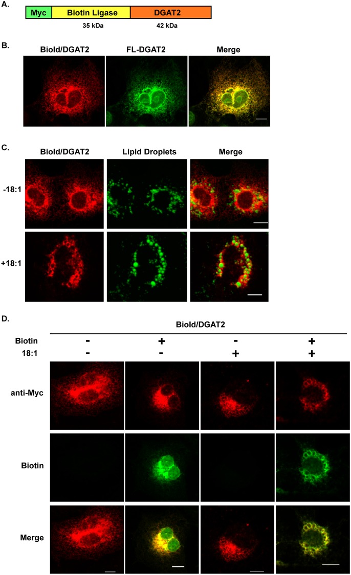
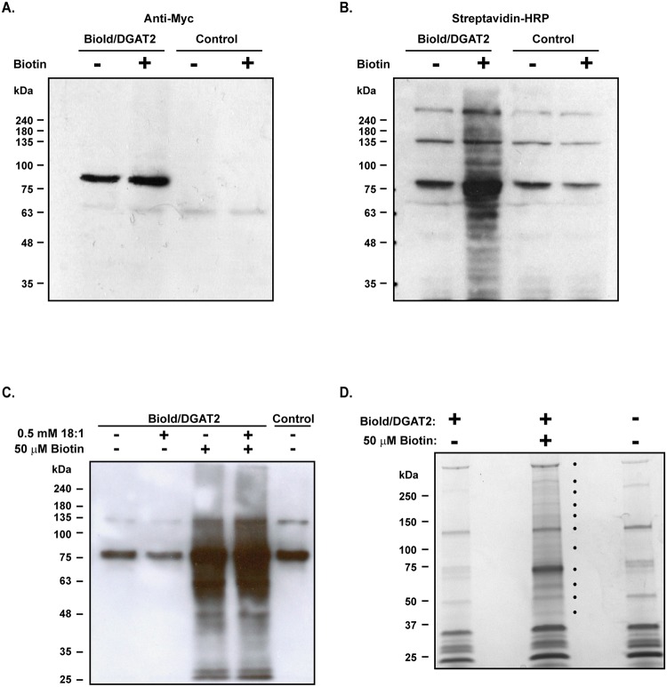
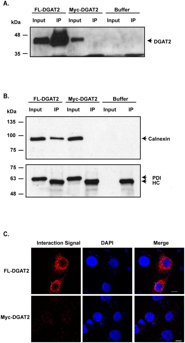
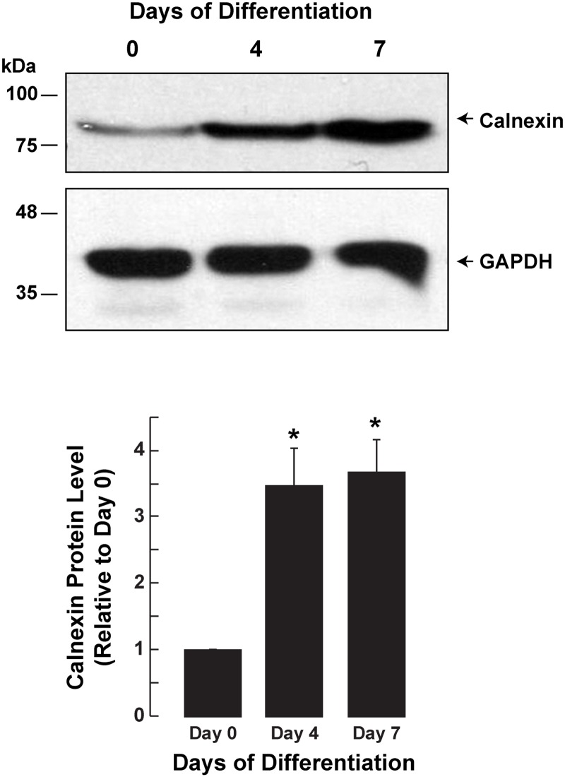
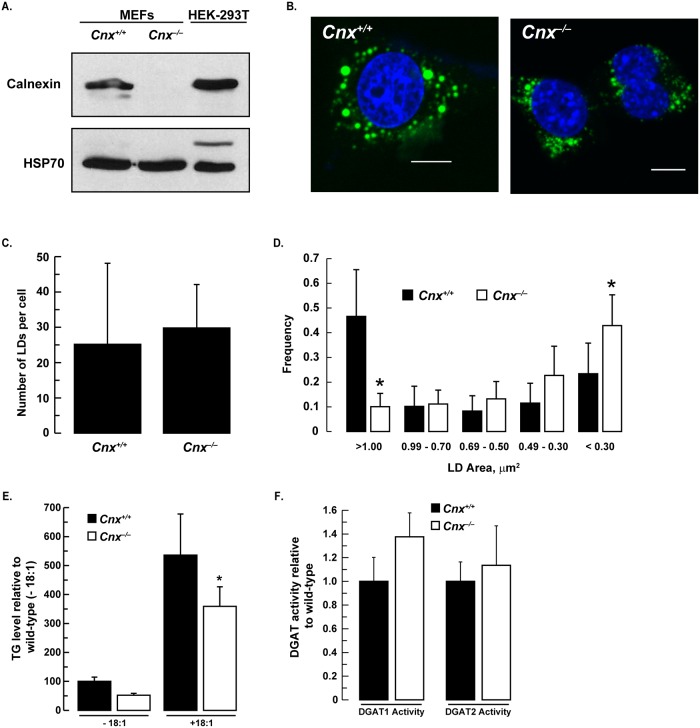
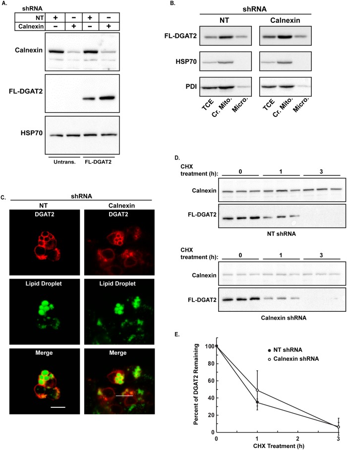
Triacylglycerol synthesis is catalyzed by acyl CoA:diacylglycerol acyltransferase-2 (DGAT2). DGAT2 is an integral membrane protein that is localized to the endoplasmic reticulum and interacts with lipid droplets. Using BioId, a method to detect proximal and interacting proteins, we identified calnexin as a DGAT2-interacting protein. Co-immunoprecipitation and proximity ligation assays confirmed this finding. We found that calnexin-deficient mouse embryonic fibroblasts had reduced intracellular triacylglycerol levels and fewer large lipid droplets (>1.0 μm2 area). Despite the alterations in triacylglycerol metabolism, in vitro DGAT2 activity, localization and protein stability were not affected by the absence of calnexin.


Conflict of interest statement

The authors have declared that no competing interests exist.

Figures

Fig 1
Fig 1. BioId/DGAT2 localizes to the ER and lipid droplets.
(A) Murine DGAT2 was fused, in frame, to the C-terminus of Myc-tagged biotin ligase. The resulting fusion protein is ~ 77 kDa. Detection of BioId/DGAT2 by immunofluorescence microscopy. (B) COS-7 cells transfected with BioId/DGAT2 and was detected with anti-Myc (red). ER was visualized by co-transfection of cells with FL-DGAT2 (green). (C) Cells expressing BioId/DGAT2 (red) were treated with or without 0.5 mM oleate for 12 h. Lipid droplets were visualized with Bodipy 493/503 (green). (D) Detection of biotinylated proteins. COS-7 cells expressing BioId/DGAT2 were cultured as described in (C) with or without 50 μM biotin for 12 h. BioId/DGAT2 was detected with anti-Myc (red) and biotinylated proteins were detected with streptavidin-488 (green). Scale bar = 10 μm.
Fig 2
Fig 2. Affinity purification of proteins biotinylated by BioId/DGAT2.
HEK-293T cells expressing BioId/DGAT2 were incubated with or without 50 μM biotin for 12 h. Cell lysates were immunoblotted with (A) anti-Myc and (B) streptavidin-HRP to detect BioId/DGAT2 and biotinylated proteins, respectively. Control: cells expressing LacZ. (C) HEK-293T cells expressing BioId/DGAT2 were incubated with or without 50 μM biotin and with or without 0.5 mM oleate for 12 h. Biotinylated proteins were captured with magnetic streptavidin beads, separated by SDS-PAGE and detected with streptavidin-HRP. (D) Separation of biotinylated proteins for in-gel digestion and mass spectrometry. Biotinylated proteins from (C) were stained with Bio-Safe Coomassie Brilliant Blue G-250 stain.
Fig 3
Fig 3. Identification of calnexin as a DGAT2 interacting protein by co-immunoprecipitation and mass spectrometry.
(A) HEK-293T cells were transfected with either FL-DGAT2 or myc-DGAT2. FL-DGAT2 was immunoprecipitated with anti-FLAG agarose from detergent solubilized material. Immunoprecipitates (IP) were separated by SDS-PAGE and were then probed with anti-DGAT2. (B) Calnexin, but not PDI, was detected in anti-FLAG immunoprecipitates by immunoblotting. HC; heavy chain. (C) Interaction of DGAT2 and calnexin was detected in situ using a proximity ligation assay. COS-7 cells expressing either FL-DGAT2 or Myc-DGAT2 were stained with mouse anti-FLAG and rabbit anti-calnexin antibodies. Interaction signals (red) were detected using a Duolink detection kit. Nuclei were stained with DAPI (blue). Scale bar = 10 μm.
Fig 4
Fig 4. Calnexin protein abundance during mouse 3T3-L1 adipocyte differentiation.
Cell lysates were prepared from 3T3-L1 cells at different days of adipocyte differentiation. Protein samples were separated by SDS-PAGE and immunoblotted for calnexin and GAPDH. Data are the mean of three experiments. *, p<0.01, Day 0 versus Days 4 and 7.
Fig 5
Fig 5. Reduced lipid droplet size in calnexin-deficient MEFs.
(A) Immunoblot showing that calnexin was not detectible in Cnx2–/– MEFs. (B) Cnx2+/+ and Cnx2–/– MEFs were incubated with 0.5 mM oleate for 12 h and then stained with Bodipy 493/503 and DAPI. Scale bars, 10 μm. Lipid droplet number (C) and area (D) were quantified using ImageJ (National Institutes of Health, rsb.info.nih.gov/ij). *, p<0.001, Cnx2+/+ versus Cnx2–/– MEFs. Mean lipid droplet number per cell and lipid droplet area were calculated from 17 to 25 cells. (E) Lipids were extracted from Cnx2+/+ and Cnx2–/– MEFs treated with or without 0.5 mM oleate for 12 h. Data are the mean of three experiments performed in duplicate. *, p<0.001, Cnx2+/+ versus Cnx2–/– oleate-loaded MEFs. (F) In vitro DGAT1 and DGAT2 activities from Cnx2+/+ and Cnx2–/– cell extracts. Data are the mean of two experiments performed in triplicate.
Fig 6
Fig 6. The subcellular localization and stability of DGAT2 is not altered in the absence of calnexin.
(A) Immunoblot with anti-calnexin showing the efficient silencing of calnexin in HEK-293T cells with shRNAs (top panel). The control (NT) refers to HEK-293T cells transduced with a non-targeting shRNA. The bottom panel shows non-targeted and calnexin knockdown cells transiently transfected with FL-DGAT2 (2 right lanes). Untransfected cells (Untrans.) are the 2 left lanes. (B) Total cell extracts (TCE), crude mitochondria (Cr. Mito.) and microsomes (Micro.) were separated by SDS-PAGE and immunoblotted with anti-FLAG, anti-PDI and HSP70 antibodies. (C) Non-targeted and calnexin knockdown cells were transfected with FL-DGAT2 and treated with 0.5 mM oleate for 12 h. After fixation and permeabilization, cells were stained with anti-FLAG and BODIPY 493/503 to visualize lipid droplets. Scale bars: 10 μm. (D) 100 μg/mL CHX was added to the culture medium of HEK-293T (non-targeted and calnexin knockdown) cells expressing FL-DGAT2. Cells were harvested 0, 1 and 3 h after the addition of CHX. The amount of FL-DGAT2 and calnexin present after CHX treatment was determined by immunoblotting. (E) Quantification of the data in Fig 6E. Data are the mean of three independent experiments, performed in triplicate.

References

    1. Cases S, Stone SJ, Zhou P, Yen E, Tow B, Lardizabal KD, et al. Cloning of DGAT2, a second mammalian diacylglycerol acyltransferase, and related family members. J Biol Chem. 2001;276:38870–6. 10.1074/jbc.M106219200 - DOI - PubMed
    1. Stone SJ, Levin MC, Zhou P, Han JY, Walther TC, Farese RV. The endoplasmic reticulum enzyme DGAT2 is found in mitochondria-associated membranes and has a mitochondrial targeting signal that promotes its association with mitochondria. J Biol Chem. 2009;284(8):5352–61. 10.1074/jbc.M805768200 - DOI - PMC - PubMed
    1. Stone SJ, Levin MC, Farese RV Jr. Membrane topology and identification of key functional amino acid residues of murine acyl-CoA:diacylglycerol acyltransferase-2. J Biol Chem. 2006;281(52):40273–82. 10.1074/jbc.M607986200 - DOI - PubMed
    1. McFie PJ, Jin Y, Banman SL, Beauchamp E, Berthiaume LG, Stone SJ. Characterization of the interaction of diacylglycerol acyltransferase-2 with the endoplasmic reticulum and lipid droplets. Biochimica et Biophysica Acta (BBA)—Molecular and Cell Biology of Lipids. 2014;1841(9):1318–28. - PubMed
    1. McFie PJ, Banman SL, Kary S, Stone SJ. Murine diacylglycerol acyltransferase-2 (DGAT2) can catalyze triacylglycerol synthesis and promote lipid droplet formation independent of its localization to the endoplasmic reticulum. J Biol Chem. 2011;286(32):28235–46. 10.1074/jbc.M111.256008 - DOI - PMC - PubMed

Publication types

Grants and funding