Review
. 2019 Mar:141:236-248.
doi: 10.1016/j.phrs.2019.01.007. Epub 2019 Jan 4.

A basic solution to activate the cholinergic anti-inflammatory pathway via the mesothelium?

Affiliations
Review

A basic solution to activate the cholinergic anti-inflammatory pathway via the mesothelium?

Elinor C Mannon et al. Pharmacol Res. 2019 Mar.

Abstract

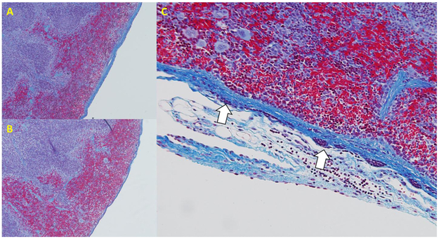
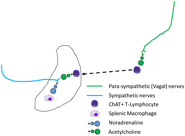
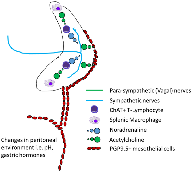
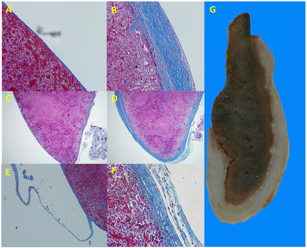
Much research now indicates that vagal nerve stimulation results in a systemic reduction in inflammatory cytokine production and an increase in anti-inflammatory cell populations that originates from the spleen. Termed the 'cholinergic anti-inflammatory pathway', therapeutic activation of this innate physiological response holds enormous promise for the treatment of inflammatory disease. Much controversy remains however, regarding the underlying physiological pathways mediating this response. This controversy is anchored in the fact that the vagal nerve itself does not innervate the spleen. Recent research from our own laboratory indicating that oral intake of sodium bicarbonate stimulates splenic anti-inflammatory pathways, and that this effect may require transmission of signals to the spleen through the mesothelium, provide new insight into the physiological pathways mediating the cholinergic anti-inflammatory pathway. In this review, we examine proposed models of the cholinergic anti-inflammatory pathway and attempt to frame our recent results in relation to these hypotheses. Following this discussion, we then provide an alternative model of the cholinergic anti-inflammatory pathway which is consistent both with our recent findings and the published literature. We then discuss experimental approaches that may be useful to delineate these hypotheses. We believe the outcome of these experiments will be critical in identifying the most appropriate methods to harness the therapeutic potential of the cholinergic anti-inflammatory pathway for the treatment of disease and may also shed light on the etiology of other pathologies, such as idiopathic fibrosis.

Keywords: Macrophage polarization; Perisplenitis; Sodium bicarbonate; Tumor-necrosis factor; Vagal nerve stimulation.


Figures

Figure 1.
Figure 1.. Cartoon demonstrating the location of PGP9.5 positive mesothelial lined connections to the rat spleen.
Solid ovals, PGP9.5 positive mesothelial cells. Open ovals, PGP9.5 negative mesothelial cells as shown in Figure 2.
Figure 2.
Figure 2.. Mesothelin and PGP9.5 localization in the spleen and heart.
Panel A) Anti-mesothelin staining of the rat cardiac atria. Mesothelial cells cover the entirety of the outer surface of the rat cardiac atria as indicated by positive staining for the mesothelial marker mesothelin (dark brown staining). Original magnification 5X, bar = 400μM. Panel B) Mesothelial cells cover the surface of the spleen and are continuous with mesothelial cells that line collagen connections that make contact with the spleen along its most anterior axis. Original magnification 5X. Panel C) Some of the mesothelial cells lining the cardiac atria stain positive for the pan-neuronal marker PGP9.5 (dark brown staining). Original magnification 5X. Panel D) higher power image of PGP9.5 positive mesothelial cells on the rat cardiac atria. Original magnification 40X, bar = 50μM. Panel E) Low power image of rat spleen stained for PGP9.5. Mesothelial cells of the diaphragmatic surface (white arrow) of the spleen are positive for PGP9.5 while those on the visceral surface are negative. Original magnification 5X.
Figure 3.
Figure 3.. Di-synaptic model of the cholinergic anti-inflammatory pathway.
Efferent parasympathetic vagal nerves (green lines) synapse with post-synaptic sympathetic fibers (blue lines) in the celiac ganglion. Nerve impulses in these post-synaptic sympathetic nerves, that enter the spleen via the splenic hilum, promote release of noradrenaline (blue circles) from nerve terminals in the splenic parenchyma which are in close proximity to Choline acetyl transferase (ChAT) positive T-lymphocytes. Stimulation of adrenergic receptors of ChAT positive T-lymphocytes by noradrenaline promotes acetylcholine (green circles) production by these cells. Increased splenic acetylcholine levels activates nicotinic acetylcholine receptors on splenic macrophages limiting the production of tumor necrosis factor α, or other pro-inflammatory cytokines, and promotes a systemic anti-inflammatory state.
Figure 4.
Figure 4.. Direct parasympathetic innervation model of the cholinergic anti-inflammatory pathway.
Vagal parasympathetic fibers (green lines) innervate the spleen at the splenic poles. Activation of efferent parasympathetic fibers directly promotes acetylcholine release within the splenic parenchyma. This pathway presumably also requires Choline acetyl transferase (ChAT) positive T-lymphocytes and splenic macrophages however specific signaling pathways within the spleen have not been detailed for this model.
Figure 5.
Figure 5.. Capsular fibrosis and capsular cell proliferation following surgical manipulation of the spleen in the mouse.
We have previously reported capsular fibrosis and mesothelial cell hypertrophy and hyperplasia on the surface of the rat spleen following surgical manipulation that disrupted thin mesothelial lined connections to the splenic capsule. While, due to the thinner capsule, fibrosis is not easily observed 3 weeks post-surgery in the mouse spleen, capsular fibrosis is evident in trichrome stained sections of the mouse spleen (A) when compared to spleens that were not manipulated during surgery (B) (original magnification of both images 5X). Panel C shows capsular thickening and cell proliferation (arrows) on the surface of the surgically manipulated mouse spleen, similar to that previously reported in the rat. Original magnification 40X.
Figure 6.
Figure 6.. A model of the cholinergic anti-inflammatory pathway initiated by stimulation of splenic sympathetic nerve terminals by circulating ChAT-positive T cells.
Vagal parasympathetic fibers (green lines) release acetylcholine (green circles) in proximity to Choline acetyl transferase (ChAT) positive T-lymphocytes at some as yet unidentified site outside the spleen. These activated ChAT positive T-lymphocytes then infiltrate the spleen via the circulation and release acetylcholine in proximity to sympathetic nerve terminals (blue line). This promotes one of either depolarization of these nerve terminals and release of noradrenaline (blue circles) or direct release of noradrenaline in the absence of terminal depolarization. This noradrenaline then stimulates β-receptors on splenic macrophages, limiting the production of tumor necrosis factor α, or other pro-inflammatory cytokines, and promoting a systemic anti-inflammatory state
Figure 7.
Figure 7.. A model of the cholinergic anti-inflammatory pathway as a gastro-intestinal - splenic axis, mediated by non-neuronal signaling through the mesothelium.
Changes in venous blood draining the GI-tract or within the peritoneal environment such as increased levels of gastric hormones or direct input from efferent vagal nerves, stimulates signaling between PGP9.5 positive mesothelial cells (red ovals). These signals reach the spleen via paracrine signaling along thin mesothelial cell lined collagen connections that adjoin the anterior edge and inferior border of the spleen and in turn activate PGP9.5 positive mesothelial cells on the capsular surface. These mesothelial cells release acetylcholine (green circles) or some other substance which stimulates release of noradrenaline or other mediators from a web of nerves underlying the capsule, derived from the sympathetic splenic nerves. This noradrenaline then stimulates β-receptors on splenic macrophages, limiting the production of tumor necrosis factor α, or other pro-inflammatory cytokines, and promoting a systemic anti-inflammatory state.
Figure 8.
Figure 8.. Progressive fibrosis and thickening of the rat splenic capsule is reminiscent of human perisplenitis and may represent a continued and unsuccessful attempt of the capsular mesothelium to reconnect signaling input.
Panel A) normal histology of the rat splenic capsule 8 weeks after surgery in which the spleen was not manipulated. Original magnification 20X. Panel B) significant thickening of the splenic capsule following manipulation of the spleen in the rat 8 weeks post-surgery. Original magnification 20X. Panel C) low power image of normal spleen and connections shown in panel A. Original magnification 5X. Panel D) low power image of thickened splenic capsule and connections in image shown in panel B. Original magnification 5X. Panel E) mesothelial cell lined collagen connection attached to the inferior border of the rat spleen in trichrome stained paraffin section. Original magnification 10X. Panel F) Example of shredding of the capsule often observed in rat with splenic capsular fibrosis, reminiscent of mesothelial lined connections present in the normal spleen. Panel G) Gross histological section of human spleen demonstrating significant splenic capsular fibrosis (perisplenitis). Note the general appearance and location of the fibrosis is similar to that observed in the rat spleen 8 weeks post-surgical manipulation (panel D). Image in panel G is modified from image shown in Monash University museum of pathology (http://museum.med.monash.edu.au/spec/index.cfm?spec=D3D1) with permission.

References

    1. Tracey KJ. The inflammatory reflex. Nature. 2002;420(6917):853–859. - PubMed
    1. Koopman FA, Chavan SS, Miljko S, et al. Vagus nerve stimulation inhibits cytokine production and attenuates disease severity in rheumatoid arthritis. Proc Natl Acad Sci U S A. 2016;113(29):8284–8289. - PMC - PubMed
    1. Kwan H, Garzoni L, Liu HL, et al. Vagus Nerve Stimulation for Treatment of Inflammation: Systematic Review of Animal Models and Clinical Studies. Bioelectron Med. 2016;3:1–6. - PMC - PubMed
    1. Ray SC, Baban B, Tucker MA, et al. Oral NaHCO3 Activates a Splenic Anti-Inflammatory Pathway: Evidence That Cholinergic Signals Are Transmitted via Mesothelial Cells. J Immunol. 2018;200(10):3568–3586. - PMC - PubMed
    1. Tracey KJ. Reflex control of immunity. Nat Rev Immunol. 2009;9(6):418–428. - PMC - PubMed

Publication types