

Noninvasive molecular diagnosis of craniopharyngioma with MRI-based radiomics approach
- PMID: 30616515
- PMCID: PMC6322318
- DOI: 10.1186/s12883-018-1216-z
Noninvasive molecular diagnosis of craniopharyngioma with MRI-based radiomics approach
Abstract
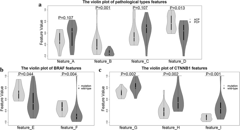
Background: Frequent somatic mutations of BRAF and CTNNB1 were identified in both histological subtypes of craniopharyngioma (adamantinomatous and papillary) which shed light on target therapy to cure this oncogenic disease. The aim of this study was to investigate the noninvasive MRI-based radiomics diagnosis to detect BRAF and CTNNB1 mutations in craniopharyngioma patients.
Methods: Forty-four patients pathologically diagnosed as adamantinomatous craniopharyngioma (ACP) or papillary craniopharyngioma (PCP) were retrospectively studied. High-throughput features were extracted from manually segmented tumors in MR images of each case. The modifications-robustness in region of interests and Random Forest-based feature selection methods were adopted to select the most significant features. Random forest classifier with 10-fold cross-validation was applied to build our radiomics model.
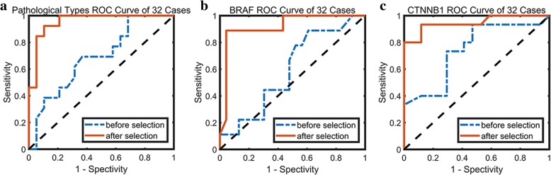
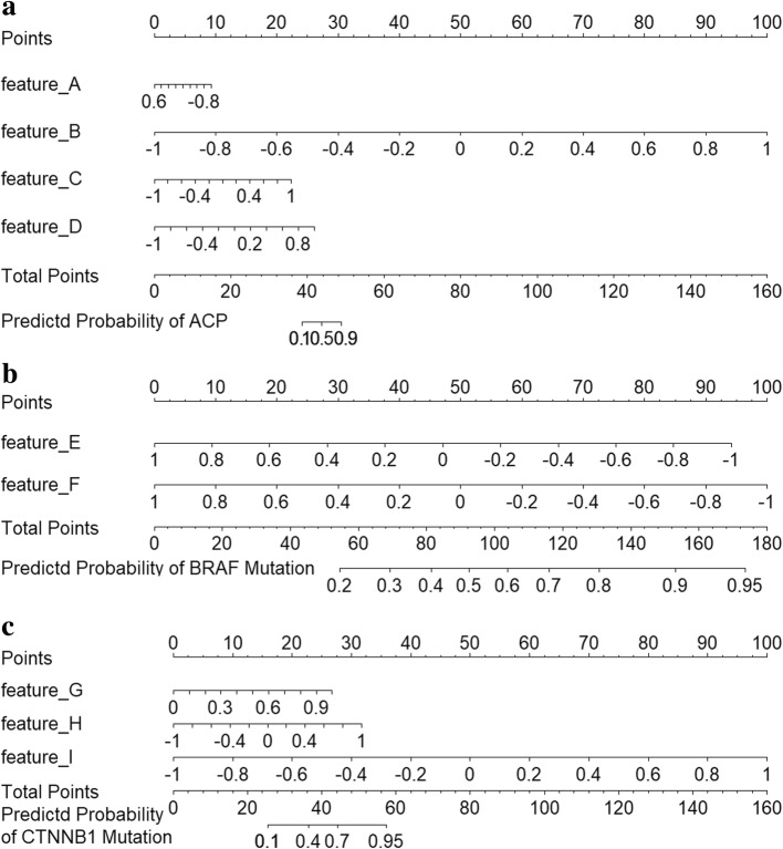
Results: Four features were selected to make pathological diagnosis between ACP and PCP with area under the receiver operating characteristic curve (AUC) of 0.89, accurancy (ACC) of 0.86, sensitivity (SENS) of 0.89 and specificity (SPEC) of 0.85. The other two features were applied to estimate BRAF V600E mutation with AUC of 0.91, ACC of 0.93, SENS of 0.83 and SPEC of 0.97. Accurate predication of CTNNB1 mutation by three selected features was realized with AUC of 0.93, ACC of 0.86, SENS of 0.86 and SPEC of 0.86.
Conclusions: We developed a reliable MRI-based radiomics approach to perform pathological and molecular diagnosis in craniopharyngioma patients with considerably accurate prediction, which could offer potential guidance for clinical decision-making.
Keywords: Craniopharyngioma; Machine learning; Molecular diagnosis; Non-invasiveness; Radiomics approach.
Conflict of interest statement
Ethics approval and consent to participate
The present study was approved by the Ethics Committee of Hushan Hospital of Fudan University. All procedures performed in studies involving human participants were in accordance with the ethical standards of the institutional and national research committee, and the 1964 Helsinki declaration and its later amendments or comparable ethical standards. A written informed consent was obtained from all individual participants included in the study. For minor patients (< 16), the written consent form was obtained from the accompanying parents.
Consent for publication
A written consent form was obtained from all participants for potentially publishing their clinical data and images while protecting their personal information. For participants under the age of 16, we obtained the written consent form from their parents.
Competing interests
The authors declare that they have no competing interests.
Publisher’s Note
Springer Nature remains neutral with regard to jurisdictional claims in published maps and institutional affiliations.
Figures


References
-
- Pascual JM, Prieto R. Harvey Cushing and pituitary case number 3 (Mary D.): the origin of this most baffling problem in neurosurgery. Neurosurg Focus. 2016;41:1. - PubMed
MeSH terms
Substances
Grants and funding
LinkOut - more resources
Full Text Sources
Medical
Research Materials
Miscellaneous
